In 2026 eastern Congo is fighting its largest Ebola outbreak on record, caused by the Bundibugyo virus, one with no approved vaccine. This film explains the fundamentals: how Ebola was found in 1976, what the thread-shaped virus is made of, how it slips into a cell using a cholesterol-carrying protein called NPC1, how it silences the body's alarm, why it spreads through contact with body fluids rather than through the air, and how contact tracing, safe burials and ring vaccination break the chain. Health information here is education, not medical advice.
Interactive 3D film9 min1976 to 2026Published 17 October 2026
Why Ebola is so deadly, and what finally stops it.
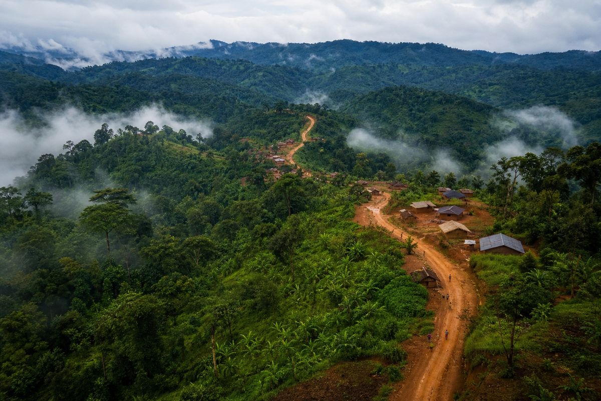
In the forests and towns of eastern Congo, health workers are fighting the largest Ebola outbreak the country has ever recorded.
By early October 2026, the World Health Organization counted more than eight thousand seven hundred confirmed cases and more than four thousand two hundred deaths.
It is caused by a rarer relative of the Ebola virus, the Bundibugyo virus, and there is no approved vaccine for it. Yet Ebola cannot travel through the air, and it cannot spread before its host feels sick. So why is it so hard to stop?
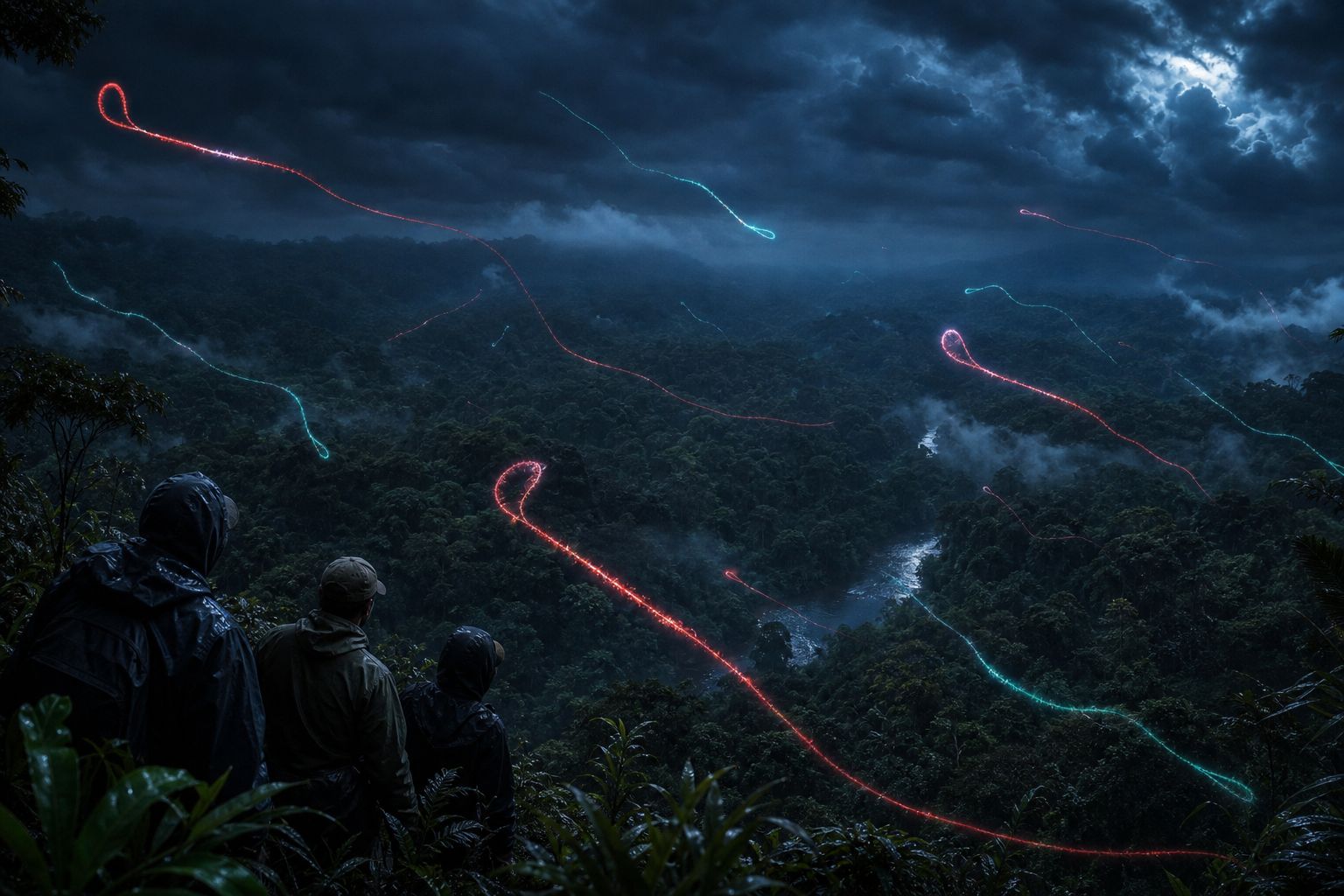
Ituri province The epicentre: forested hills on Congo’s border with Uganda. AI-generated still.
8,728
cases in Congo
4,205
deaths
48%
of confirmed cases died
Confirmed, to 6 October 2026 (WHO)
first identified in Uganda, 2007
no approved vaccine or specific treatment (WHO)
7 of Congo’s 26 provinces affected
Bundibugyo virus
Prologue · The news
Congo’s largest Ebola outbreak
Congo confirmed the outbreak in mid-May 2026. WHO’s Disease Outbreak News of 8 October 2026 reported 8,728 confirmed cases and 4,205 confirmed deaths in 64 health zones in seven provinces, with Ituri the most affected. WHO calls it the largest Ebola outbreak ever recorded in the country, whatever the virus species. According to CDC, Uganda, Kenya and France have reported a small number of linked cases.
For comparison, the 2014–2016 West African epidemic, caused by a different species, killed 11,325 people.
This film is education, not medical advice. Anyone who may have been exposed should contact local health authorities.
How to use: pick a chapter or press ← →; drag to rotate. Many steps have buttons you can press.
Two outbreaks in the same year, hundreds of kilometres apart.
Ebola was first recognised in 1976, in two separate outbreaks. One began in Nzara, in what is now South Sudan, caused by a closely related virus now called the Sudan virus. The other began weeks later at a mission hospital in Yambuku, in what is now the Democratic Republic of the Congo.
At Yambuku, three hundred and eighteen people fell ill and two hundred and eighty died. Investigators found that needles reused without proper sterilising had carried the virus from patient to patient.
The new virus was named after the Ebola River, near the village.
first Nzara, Sudan: 284 cases, 151 deaths
Aug–Nov Yambuku, Zaire: 318 cases, 280 deaths
1976
Nzara: Sudan virus
Yambuku: Ebola virus
Two different viruses
88%
of those who fell ill died
1 hospital
needles rinsed in warm water between patients
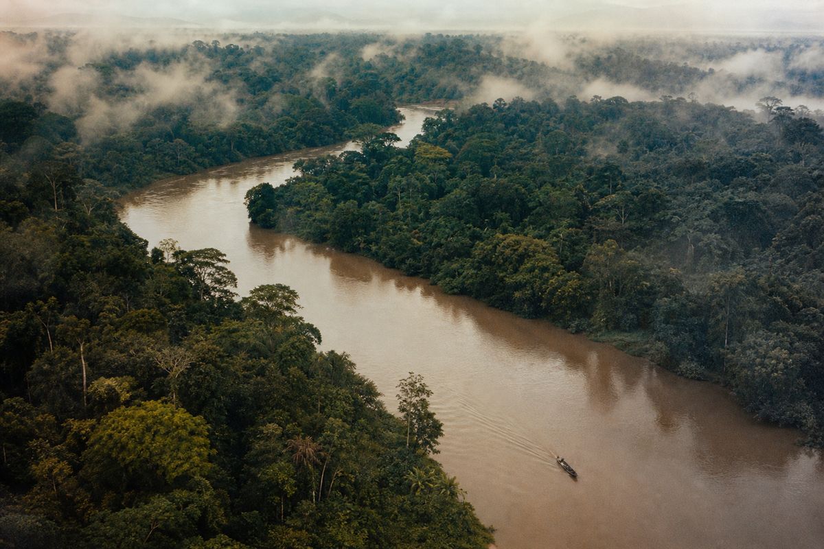
Yambuku, 1976A rainforest river The virus took the name of a nearby river, not of the village. AI-generated still.
Chapter I · The thread
The first outbreaks
WHO dates the first two outbreaks to 1976: Sudan virus disease in Nzara (now South Sudan) and Ebola virus disease in Yambuku (now the DRC), near the Ebola River. At Yambuku Mission Hospital, 318 people fell ill and 280 died. The investigation, which included the Belgian microbiologist Peter Piot, traced most early infections to vitamin injections given with needles that were rinsed in warm water rather than sterilised.
The Sudan outbreak began a few weeks earlier and was caused by a different, related virus.
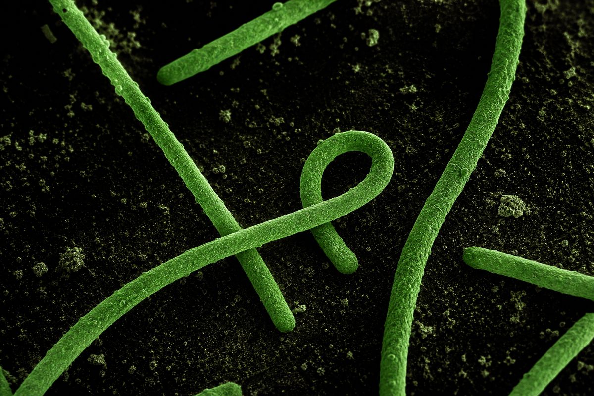
Under an electron microscope, Ebola looks like a thread, sometimes curled into a loop at one end. It belongs to the filoviruses, from the Latin word for thread.
It is about eighty nanometres wide and roughly a thousand long. Inside is a single strand of RNA carrying just seven genes.
On the outside, spikes made of one protein, the glycoprotein, stud the surface. They are the keys the virus uses to get into our cells.
Thread viruses Coloured micrographs often show the “shepherd’s crook” curl. AI-generated illustration.
~80 nm
wide: a thousandth of a hair
~19,000
letters of RNA
7
genes
Ebola virus (ViralZone)
GP: the surface spike
NP, VP30, VP35, L: copy the RNA
VP40, VP24: build the shell and bud out
Its seven genes make
Chapter I · The thread
What the virus is
ViralZone describes the Ebola virion as filamentous, about 80 nm in diameter and around 970 nm long, enclosing a linear, negative-sense RNA genome of about 18–19 kilobases that encodes seven structural proteins: NP, VP35, VP40, GP, VP30, VP24 and the polymerase L. The glycoprotein GP forms the spikes used for attachment and entry.
The 3D virus and cell are schematic models, not to scale.
Scientists now know six species of these viruses. Three have caused large human outbreaks: the original Ebola virus, the Sudan virus, and the Bundibugyo virus, first found in Uganda in 2007.
They are close cousins, but different enough that a vaccine or drug built against one may not work against another.
Ebola virus: Ebola virus disease
Sudan virus: Sudan virus disease
Bundibugyo virus: Bundibugyo virus disease
Large human outbreaks (WHO)
Bundibugyo, Uganda 2007
~30%
Bundibugyo, Congo 2012
~50%
Bundibugyo, Congo 2026
48%
Ebola virus, Yambuku 1976
88%
Share of patients who died, by outbreak
Ebola River, Congo
Taï Forest, Côte d’Ivoire
Bundibugyo district, Uganda
Reston, Virginia, USA
Named after where they were found
Chapter I · The thread
Six orthoebolaviruses
WHO lists six species in the genus Orthoebolavirus (family Filoviridae). Three cause large outbreaks: Ebola virus (EBOV), Sudan virus (SUDV) and Bundibugyo virus (BDBV). The approved vaccine Ervebo targets EBOV only. WHO gives case fatality of about 30% for the 2007 Bundibugyo outbreak in Uganda and about 50% for the 2012 one in Congo.
Fatality varies with the virus, the care available and how quickly patients reach it.
A virus cannot do anything on its own. To make copies, it must get inside a living cell and hijack it.
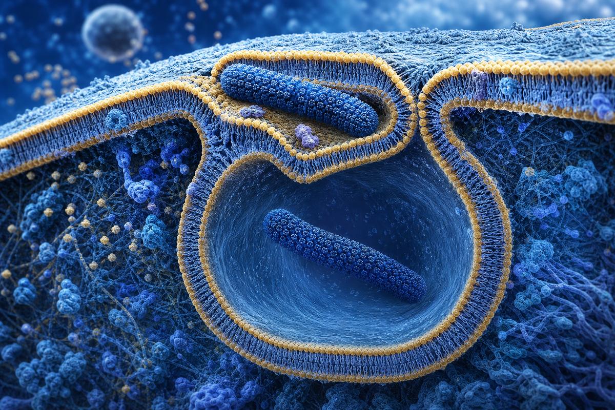
Ebola's spikes stick to molecules on the surface of a cell. The cell responds by doing something it does every day: it folds its membrane around the particle and gulps it into a bubble.
This gulping is called macropinocytosis. The virus is now inside, but still trapped in the bubble.
raw materials to build copies
the cell’s protein factories
energy
What a virus needs from a cellGulped into a bubble The membrane ruffles and closes around the particle. Schematic, AI-generated illustration.
macro + pino + cytosis
Greek for “big drinking by the cell”
Step 1 of the cycle
Chapter II · Breaking in
Attachment and uptake
According to ViralZone, the glycoprotein binds attachment factors on the cell surface (such as the lectins DC-SIGN and the receptor TIM-1, which recognises a lipid on the viral envelope). This triggers macropinocytosis, and the virion is carried into a large internal vesicle, the macropinosome.
The 3D virus and cell are schematic models, not to scale.
Inside the bubble, the cell's own enzymes trim the virus's spikes. That exposes a hidden tip, which fits a protein in the bubble's wall called NPC1.
NPC1's everyday job is to move cholesterol. In 2011, two teams showed that Ebola cannot infect a cell without it. Cells lacking NPC1 resisted infection.
Once the spike grips NPC1, the virus's envelope fuses with the bubble wall, and its RNA spills into the cell. Try removing NPC1 yourself.
cell enzymes (cathepsins) trim the spike
the trimmed spike binds NPC1
membranes fuse; the RNA is released
Unlocking, in order
If cells don’t make NPC1, they cannot be infected by Ebola virus.ScienceDaily, on Carette et al. and Côté et al., Nature (2011)
cholesterol
carried out of the bubble into the cell
NPC1’s day job
NPC1 partly missing
most survived a lethal dose of Ebola
In mice (2011)
Chapter II · Breaking in
NPC1, the intracellular receptor
Two papers in Nature in 2011 (Carette et al.; Côté et al.) showed that Niemann–Pick C1, a cholesterol transporter in the membranes of late endosomes, is essential for Ebola virus entry. Cells without NPC1 resisted infection, mice partly lacking it mostly survived a lethal challenge, and small molecules that block NPC1 inhibited infection. ViralZone summarises the sequence: cathepsins cleave GP inside the vesicle, the cleaved GP binds NPC1, and fusion releases the genome.
These were laboratory results. No NPC1-blocking drug is an approved Ebola treatment.
The 3D virus and cell are schematic models, not to scale.
Now the virus's own enzyme reads its seven genes, and the cell's machinery builds viral proteins and fresh copies of the RNA.
The new pieces gather under the cell's outer membrane and push out through it, wrapping themselves in a piece of it as they go. Each one buds off as a new thread, ready to infect the next cell.
L, the viral polymerase, reads the genes one by one
the cell’s ribosomes make the proteins
NP wraps each new RNA copy
Inside the cell
VP40 lines the membrane from inside
the cell’s own ESCRT machinery pinches each bud off
the envelope is borrowed from the host
Budding
cytoplasm
Ebola copies itself outside the nucleus
Where it all happens
Chapter II · Breaking in
Replication and budding
Ebola replicates in the cytoplasm. The viral polymerase L transcribes the genes in sequence; once enough nucleoprotein is made, new genomes are copied and wrapped. The matrix protein VP40 brings the nucleocapsid to the plasma membrane, and new virions bud off using the host cell’s ESCRT complexes.
The 3D virus and cell are schematic models, not to scale.
Ebola attacks the very cells meant to raise the alarm.
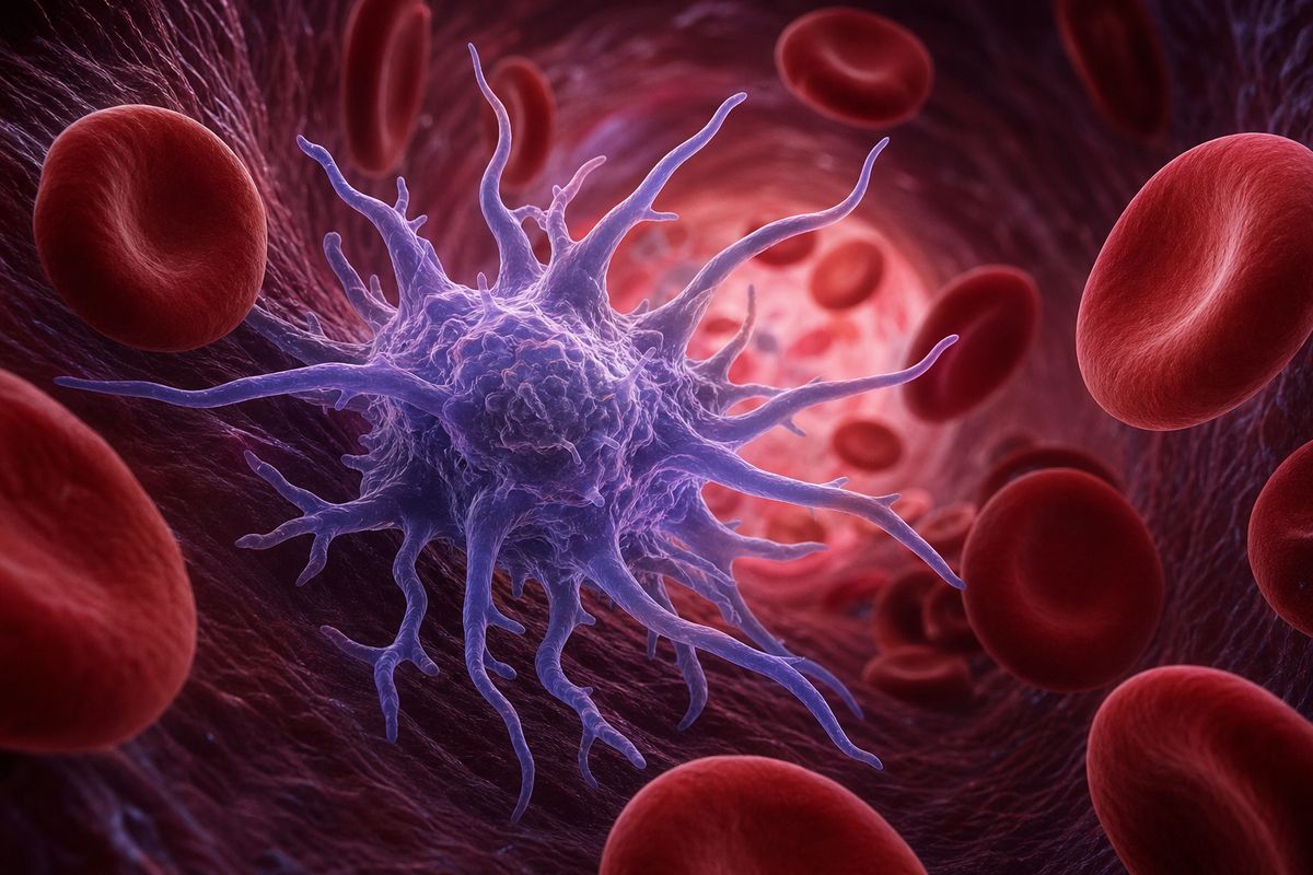
Many viruses must first get past the immune system. Ebola goes straight for it. Among its first targets are macrophages and dendritic cells, the sentries that normally sound the alarm.
An infected cell usually releases interferon, a chemical alarm that tells neighbouring cells to shut their doors. Two Ebola proteins, VP35 and VP24, jam that signal.
With the alarm silenced, the virus multiplies freely, and the infected sentries carry it through the body. Press the buttons to compare.
A sentry cell Dendritic cells reach out with long arms to sample their surroundings. AI-generated illustration.
VP35: stops the cell from sensing viral RNA
VP24: stops the interferon message from reaching the nucleus
Two saboteurs
type I interferon
the defence Ebola suppresses most (review, 2015)
The alarm
high levels of virus in the blood
infected sentries spread it to liver, spleen and other organs
the immune response comes late and out of balance
Then
Chapter III · In the body
Immune evasion
A 2015 review in Nature Reviews Microbiology describes filovirus disease as driven by potent suppression of the type I interferon response. Ebola VP35 blocks signalling through RIG-I-like receptors, the sensors that detect viral RNA, and VP24 blocks the nuclear import of the transcription factor STAT1, so interferon cannot switch on antiviral genes. Macrophages, monocytes and dendritic cells are major targets.
Bray and Geisbert (2005) describe infected macrophages releasing inflammatory signals that contribute to leaky blood vessels and clotting problems.
The 3D virus and cell are schematic models, not to scale.
Symptoms appear between two and twenty-one days after infection. At first they look like many other illnesses: fever, aches, headache and weakness.
After a few days, vomiting and diarrhoea can drain the body of fluid, and some patients bleed. An overwhelmed immune system and leaky blood vessels can lead to shock, a dangerous fall in blood pressure, and to organ failure.
On average about half of patients have died in past outbreaks. But early supportive care, replacing fluids and treating symptoms, improves survival.
fever, intense headache
muscle and joint aches
weakness, sore throat
Early, “dry” symptoms (CDC)
vomiting, diarrhoea, stomach pain
unexplained bleeding, in some patients
usually after 4–5 days of illness
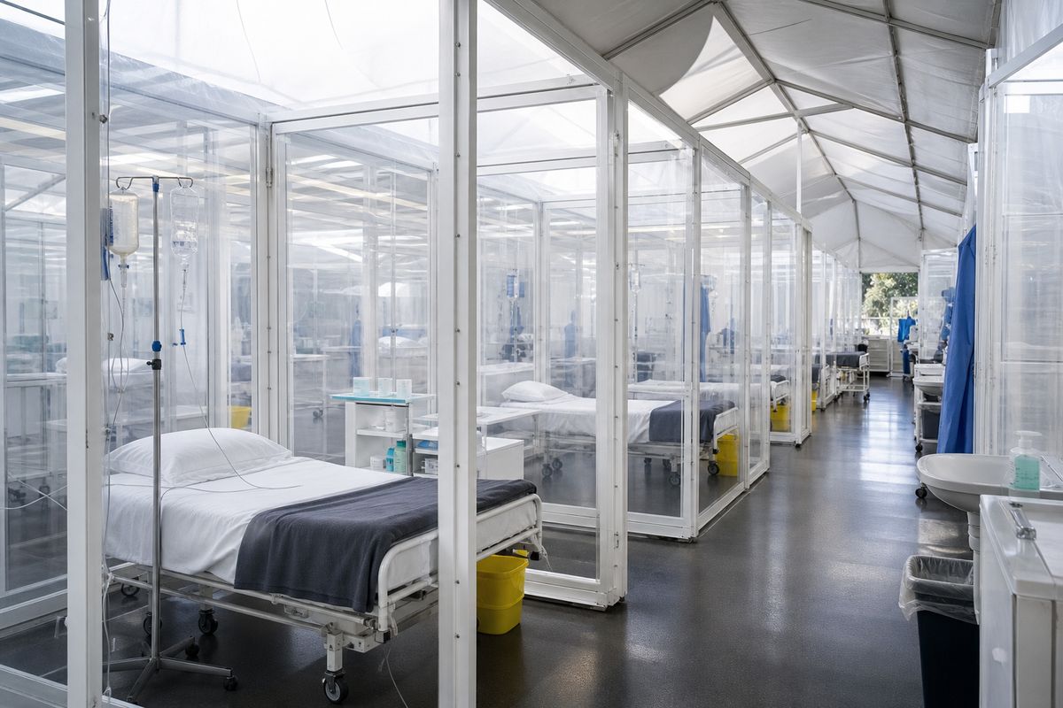
Later, “wet” symptoms (CDC)A treatment unit Each patient has a separate space, with fluids given through a drip. AI-generated still.
~50%
average
25–90%
range between outbreaks
Fatality in past outbreaks (WHO)
Chapter III · In the body
The course of the disease
CDC lists early “dry” symptoms (fever, aches, headache, weakness, sore throat) and later “wet” ones after four to five days of illness: vomiting, diarrhoea, abdominal pain and, in some patients, unexplained bleeding. WHO gives an average case fatality of about 50%, ranging from 25% to 90% in past outbreaks, and notes that early supportive care with rehydration and symptomatic treatment improves survival. For Ebola virus disease (not Bundibugyo), WHO recommends two monoclonal antibody treatments.
This film is education, not medical advice. Anyone who may have been exposed should contact local health authorities.
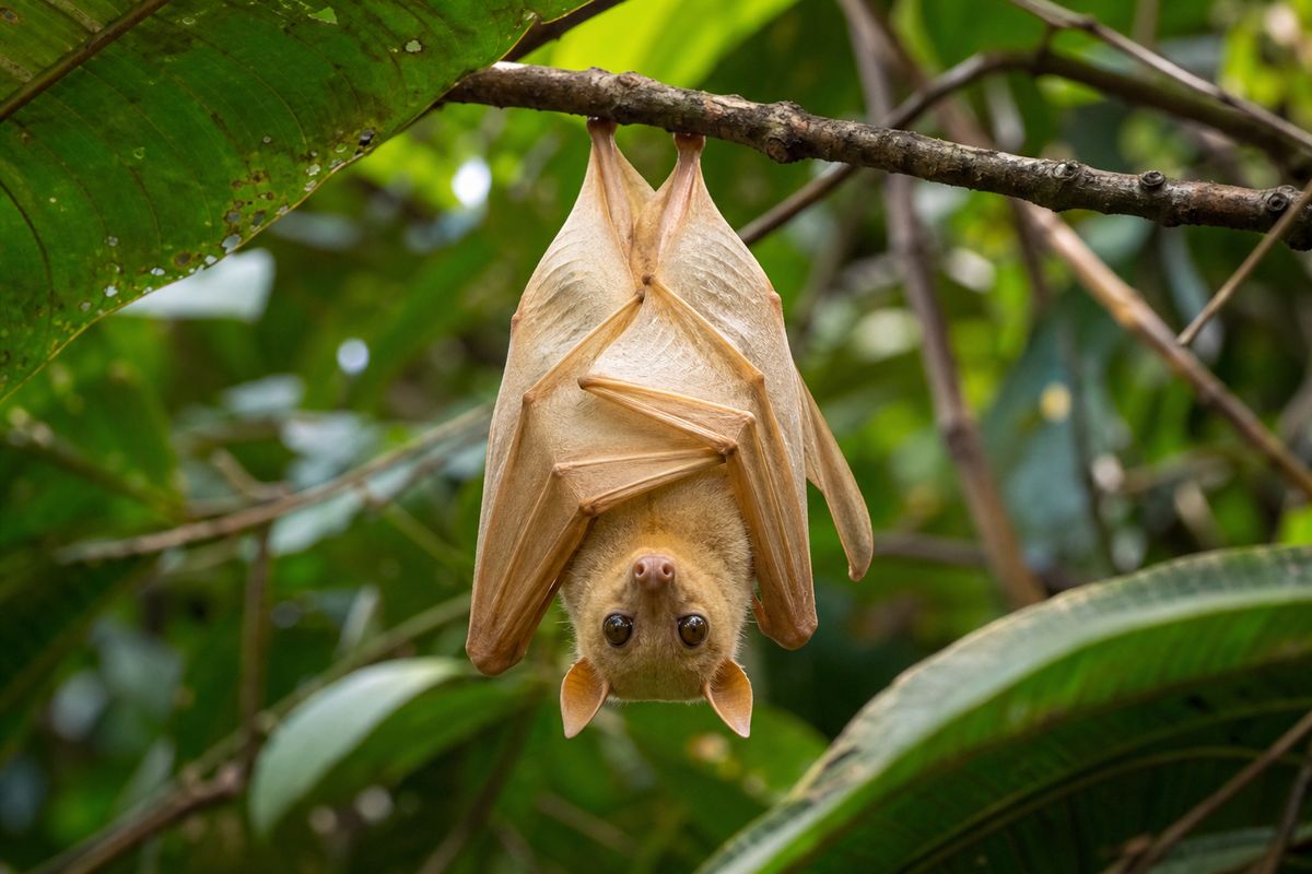
Between outbreaks, the virus survives somewhere in the wild. Fruit bats are thought to be its natural hosts, though this has not been fully proven.
People can catch it by handling infected animals: bats, but also chimpanzees, gorillas, monkeys and forest antelope. One such jump from an animal can start a human chain.
That is why outbreaks have mostly begun in or near the rainforests of central and west Africa.
Fruit bats The suspected hosts belong to the family Pteropodidae, the fruit bats. AI-generated still.
fruit bats
chimpanzees, gorillas, monkeys
forest antelope, porcupines
Animals linked to human infections (WHO)
1976 Yambuku and Nzara
2007 Bundibugyo, Uganda
2014–16 Guinea, Liberia, Sierra Leone
2026 Ituri and beyond, Congo
Some outbreaks on the map
Chapter IV · How it spreads
Animal hosts
WHO: fruit bats of the Pteropodidae family are thought to be natural Ebola virus hosts. The virus is introduced into the human population through close contact with the blood, secretions, organs or other body fluids of infected animals such as fruit bats, chimpanzees, gorillas, monkeys, forest antelope or porcupines found ill or dead or in the rainforest. WHO’s Bundibugyo update calls the bat reservoir “suspected”.
Ebola spreads through body fluids, from the sick and the dead.
Ebola does not spread through the air like flu or measles. It passes through direct contact with the blood or other body fluids of a sick person, through broken skin or the lining of the eyes, nose and mouth.
People do not spread it before they have symptoms. That is good news. But the sicker a patient becomes, the more virus they carry, so the people most at risk are those who care for them: family members, and health workers without protection.
And the virus remains in the body after death. Funerals where mourners wash or touch the body have spread it again and again.
blood, vomit, faeces and other body fluids
bedding, clothes or needles soiled with them
not through the air between people
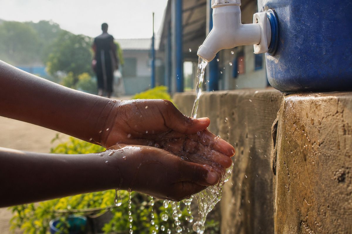
How it passes (WHO)Chlorinated water Handwashing stations stand at clinic and village entrances during outbreaks. AI-generated still.
family members caring for the sick
health workers without protection
mourners who touch the body
Most at risk (WHO)
264 of 372
reported deaths were in the community, not in a treatment centre
Congo, deaths since WHO’s previous update (8 Oct)
Chapter IV · How it spreads
Transmission
WHO: human-to-human transmission is by direct contact, through broken skin or mucous membranes, with the blood or body fluids of a person who is sick with or has died from the disease, or with objects contaminated with these fluids. People cannot transmit the disease before symptoms appear, and remain infectious as long as their blood contains the virus. Health workers are frequently infected when infection control is lax, and burials that involve direct contact with the body can spread infection.
WHO’s 8 October update reports that of 372 deaths since its previous update, 264 happened in the community rather than in treatment centres, which means more unprotected contact with patients and bodies.
The outbreak simulation is a toy model to show the logic of transmission; it does not forecast any real outbreak.
Above one, an outbreak grows. Below one, it dies out.
Epidemiologists track one number: how many people, on average, each patient goes on to infect. They call it R.
Early in the West African epidemic of 2014, R was estimated at between about 1.7 and 2. For a disease that kills so many, that may sound low.
But any value above one means each new round of cases, the people infected by the round before, is bigger than the last. Push R below one, and the chain shrinks until it breaks. Try both.
casesn+1≈R×casesn
R
average new infections per case
n
round of cases
Guinea
1.71
Liberia
1.83
Sierra Leone
2.02
Estimated R early in 2014 (NEJM)
× 58
cases at R = 1.5
× 0.03
cases at R = 0.7
Ten rounds later
R below 1
and kept there until the last case
The goal of every response
Chapter IV · How it spreads
The reproduction number
The WHO Ebola Response Team (NEJM, September 2014) estimated the basic reproduction number as 1.71 in Guinea, 1.83 in Liberia and 2.02 in Sierra Leone, with a mean incubation period of 11.4 days. The 2014–2016 epidemic eventually caused 28,652 cases and 11,325 deaths (CDC).
The “ten rounds” figures are simple arithmetic: 1.5¹⁰ ≈ 58 and 0.7¹⁰ ≈ 0.03.
The outbreak simulation is a toy model to show the logic of transmission; it does not forecast any real outbreak.
Because Ebola only spreads from people who are already sick, the oldest tools still work. Teams list everyone a patient has touched and check on each of them for twenty-one days, the upper end of the incubation period.
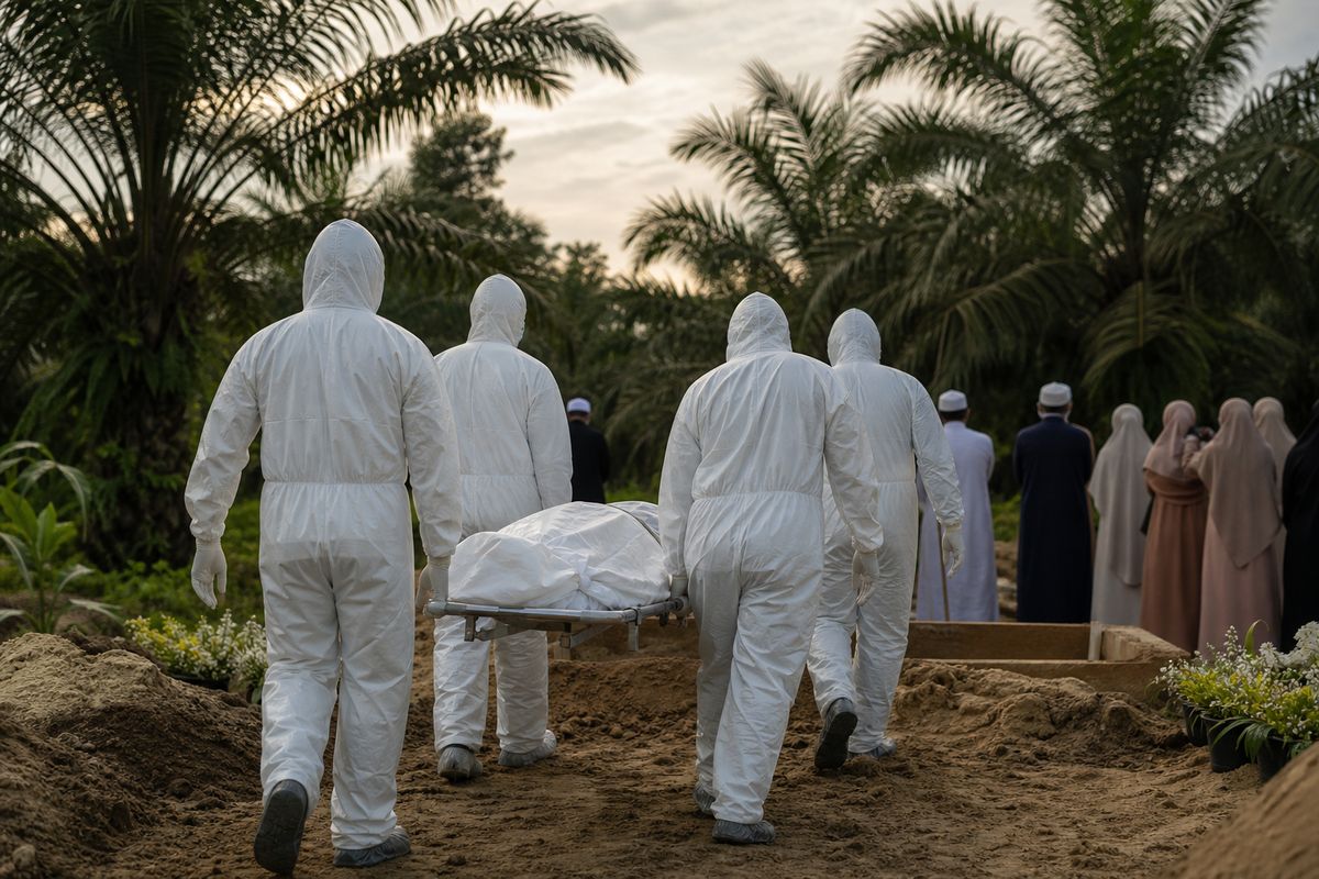
Anyone who develops a fever is isolated and cared for before they can infect others. And burial teams in protective suits help families bury their dead safely and with dignity.
Every contact traced is a branch cut from the chain. Switch tracing on and watch R fall.
find and follow contacts for 21 days
isolate and treat the sick early
infection control in clinics
work with communities
The basic toolkit (WHO)
21 days
the upper end of the incubation period (WHO: 2–21 days)
The follow-up windowSafe and dignified burial Teams wear full protection; families can take part from a safe distance. AI-generated still.
80.4%
of contacts followed up
85%
the target
Congo, latest report (WHO)
Chapter V · Breaking the chain
Tracing, isolation, safe burials
WHO’s list of control measures: contact tracing (finding people exposed and monitoring them for 21 days), separating the sick from the healthy and isolating patients in treatment centres, safe and dignified burials, infection prevention and control in health facilities, surveillance and laboratory testing, and community engagement. In its 8 October 2026 update WHO reported contact follow-up at 80.4%, below the 85% target.
The outbreak simulation is a toy model to show the logic of transmission; it does not forecast any real outbreak.
Vaccinate the contacts, and the contacts of contacts.
In 2015, in Guinea, a vaccine against the original Ebola virus was tested in a new way. Instead of vaccinating everyone, teams vaccinated a ring around each new case: the contacts, and the contacts of those contacts.
Some rings were vaccinated at once, others three weeks later. Among those vaccinated at once, no one developed Ebola from ten days after vaccination.
That vaccine, now called Ervebo, was approved in 2019. A ring of immunity around each case starves the virus of new people to infect. Switch the rings on and off and compare.
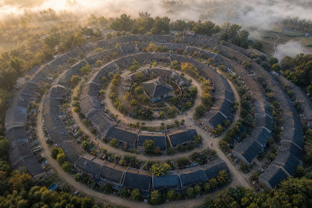
Rings of households A ring is a social circle, not a fixed distance: who visited, who cared, who lives nearby. AI-generated still.
0
cases among 2,119 vaccinated at once
16
cases in the delayed rings
100%
estimated efficacy (statistical range 69–100%)
Ebola ça Suffit! trial (The Lancet, 2017)
2015 ring trial in Guinea
2017 final results, The Lancet
Dec 2019 approved in the US (FDA)
From trial to approval
one dose
protects against Ebola virus (Zaire) only
not approved for Sudan or Bundibugyo viruses
Ervebo
Chapter V · Breaking the chain
Ring vaccination
In the Ebola ça Suffit! trial in Guinea and Sierra Leone, rings of contacts and contacts of contacts were randomised to immediate or 21-day-delayed vaccination with one dose of rVSV-ZEBOV. There were no cases with onset 10 or more days after randomisation among the 2,119 people vaccinated immediately, against 16 cases in the delayed clusters: an estimated efficacy of 100% (95% CI 68.9–100) (Henao-Restrepo et al., The Lancet, 2017). The vaccine, Ervebo, targets Ebola virus only (WHO).
The US FDA approved Ervebo on 19 December 2019.
The trial was open-label (participants knew whether they had been vaccinated), and the confidence interval is wide; the result has been borne out in later outbreaks where the vaccine was used.
The outbreak simulation is a toy model to show the logic of transmission; it does not forecast any real outbreak.
Ervebo and the antibody treatments were built against the spike of the original Ebola virus. Bundibugyo's spike is different, so for this outbreak there is no approved vaccine or specific drug.
Scientists are choosing the most promising candidate vaccines, and a treatment trial began in July 2026. Until results arrive, the chain must be broken the old way: tracing, isolation, care and safe burials.
Ebola virus: 2 vaccines, 2 antibody treatments
Sudan virus: none yet
Bundibugyo virus: none yet
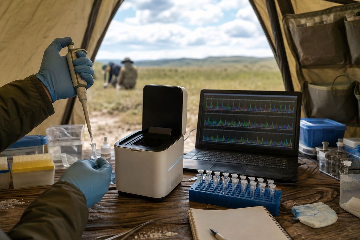
Approved tools, by virus (WHO)Field genomics Sequencing virus samples shows how cases are linked, and whether the virus is changing. AI-generated still.
tracing and isolation
early supportive care
safe burials
community trust
Until then
Chapter V · Breaking the chain
Where things stand
WHO’s 8 October 2026 update states that no approved vaccines or specific treatments exist for Bundibugyo virus disease, refers to a WHO review prioritising candidate vaccines, and notes that a clinical trial of treatments began enrolling patients in July 2026. It reports no results yet. The response relies on surveillance, testing, infection prevention, clinical care, community engagement and safe burials.
This film is education, not medical advice. Anyone who may have been exposed should contact local health authorities.
A virus with seven genes, and a chain made of people.
In Ituri, the outbreak is not over. Fifty years after Yambuku, the virus is still a thread of RNA with seven genes, which needs a lock inside our cells and silences our first alarm.
But it can only travel by touch, from the sick and the dead to the people who care for them. That is its weakness.
Every earlier outbreak has ended, and each ended the same way: by finding contacts and closing the ring.
1976 first outbreaks: Yambuku and Nzara
2007 Bundibugyo virus identified, Uganda
2011 NPC1 found to be the lock Ebola needs
2014–16 West Africa: 11,325 deaths
2015 ring vaccination trial, Guinea
2019 Ervebo approved
2026 Bundibugyo outbreak, Congo
Fifty yearsBy touch All stills and illustrations in this film are AI-generated.
find and follow every contact
isolate and care for the sick early
safe, dignified burials
a vaccine ring, where a vaccine exists
What breaks the chain
Epilogue · Sources
How Ebola works
The biology in this film (the virus’s structure, NPC1-dependent entry, interferon suppression) and the epidemiology (contact transmission, no spread before symptoms, the 21-day window, ring vaccination) are well established and summarised by WHO, CDC and peer-reviewed reviews. Figures for the 2026 outbreak are WHO’s as of 6 October 2026 and will change.
The 3D virus, cell and outbreak simulation are schematic teaching models, not to scale and not forecasts. Map pins are approximate. All stills are AI-generated. This film is education, not medical advice.