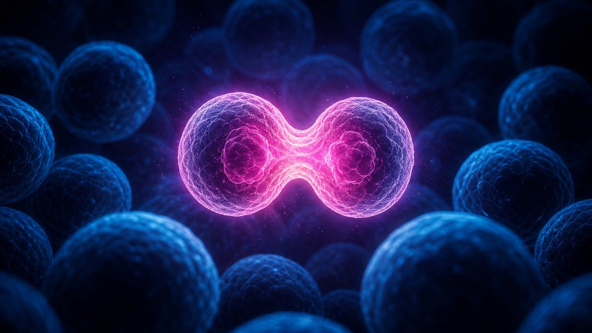
A new analysis reports several cancers rising among people under fifty, even though most cancers still strike older people. To see why age matters so much, this film follows one cell: how the body balances division and death, how DNA copying errors and carcinogens create mutations, why cancer needs both a stuck accelerator and broken brakes, what the guardian protein p53 does, why hits pile up with age, and the tricks a tumour needs to become dangerous: immortality, its own blood supply and the ability to spread. Seed a rogue cell, break p53, run Knudson’s two-hit lives and switch on telomerase yourself. Education, not medical advice.
Interactive 3D film9 min1961 to 2026Published 16 October 2026
Cancer is mostly a disease of older age. Yet in October 2026, the World Cancer Research Fund reported that several cancers are rising among people under fifty in England, the United States and the Netherlands.
Early-onset cancer is still uncommon, and the researchers say its causes are not fully understood. But it raises a question: why does age matter so much in the first place?
To answer that, we have to go inside the body, to a single cell that stops taking orders.
3.9 → 8.4
bowel cancer per 100,000 under-50s, 2001 → 2023
7
cancer types with significant rises
Early-onset cancer, England
obesity, diet, inactivity, alcohol, smoking
possibly environmental exposures
partly better awareness and earlier diagnosis
lung cancer in under-50s is falling
What the researchers point toMostly a disease of age The median age at diagnosis is above 60. AI-generated still.
Prologue · The news
Cancer in younger adults
A World Cancer Research Fund analysis published in October 2026 found significant rises in early-onset cancer (diagnosed under 50) for seven cancer types in England, five in the US and four in the Netherlands. In England, bowel cancer in under-50s rose from 3.9 to 8.4 per 100,000 between 2001 and 2023.
The researchers say causes are not fully understood; better detection may explain part of the rise but, they say, not all of it. This film explains how cancer starts. It is education, not medical advice: questions about your own health belong with a doctor.
How to use: pick a chapter or press ← →; drag to rotate. Ten steps have buttons you can press.
A body is a society of cells that follow the rules.
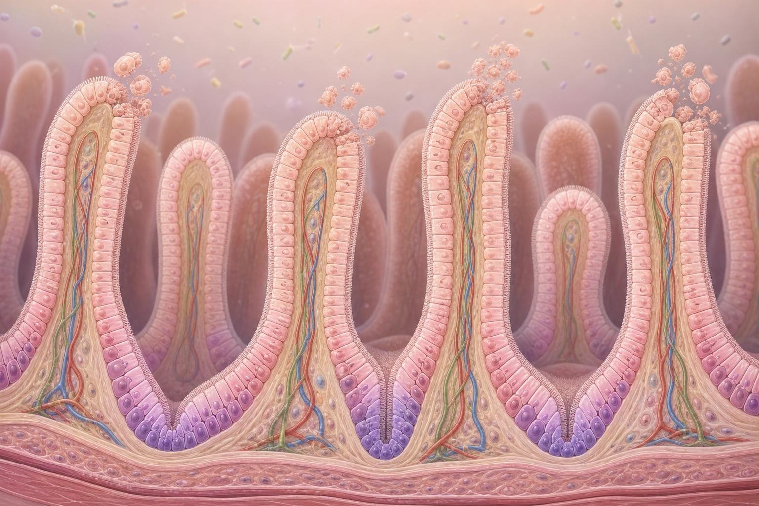
Your body holds about thirty trillion human cells. Many of them wear out, and every day around fifty to seventy billion are retired on purpose, in a tidy process called apoptosis.
Neighbours divide to fill the gaps. When a wound has healed, they slow down again. Each cell divides only when the body asks, and stops when it is told.
That is the bargain that keeps a body in shape. Cancer begins when one cell breaks it.
~30 trillion
human cells (Sender, Fuchs & Milo, 2016)
50–70 billion
cells lost to apoptosis per day
Every dayConstant renewal Linings like the gut are rebuilt continually: new cells form below as old ones are shed. AI-generated illustration.
divide only on a signal
stop when crowded
self-destruct if badly damaged
stay in your own tissue
The rules
Chapter I · The bargain
Cell turnover
A 2016 study by Sender, Fuchs and Milo estimated about 30 trillion human cells in a body. The average adult loses roughly 50 to 70 billion cells a day to apoptosis, a regulated form of cell death. Normal cells divide to replace worn-out or injured cells and return to their usual rate once a wound heals.
The 3D models are schematic and use illustrative numbers.
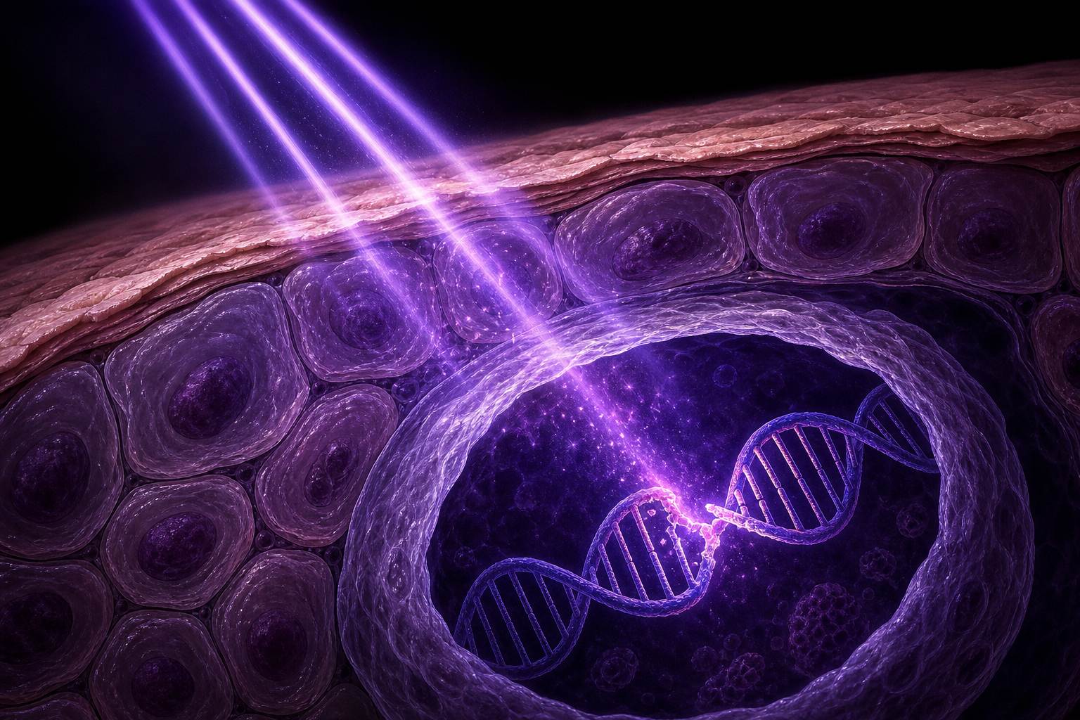
The rules are written in DNA, and every time a cell divides it must copy all of it. Cells check and repair their copies, but now and then a mistake slips through. That lasting change is a mutation.
According to the US National Cancer Institute, most cancers come from changes like these that pile up by chance over many years. Some are speeded up by carcinogens: tobacco smoke, ultraviolet light, certain viruses.
Only up to one in ten cancers is linked to a change inherited from a parent.
random copying mistakes
damage from carcinogens
inherited from a parent (rarer)
Where mutations come fromUltraviolet damage UV light can kink a DNA strand; if it is copied before it is repaired, the error becomes permanent. AI-generated illustration.
up to 10%
of cancers may be caused by inherited genetic changes (NCI)
Inherited cancer risk
Chapter I · The bargain
Cancer is a genetic disease
The National Cancer Institute calls cancer a genetic disease: it is caused by changes to genes that control how cells grow and multiply. These arise from random mistakes during cell division, from carcinogens such as tobacco smoke, UV rays and HPV, or are inherited. “The vast majority of cancers occur by chance”; up to 10% may be caused by inherited changes.
The 3D models are schematic and use illustrative numbers.
Which mistakes matter? Think of a cell as a car. Some genes are the accelerator: they push the cell to divide when growth is needed.
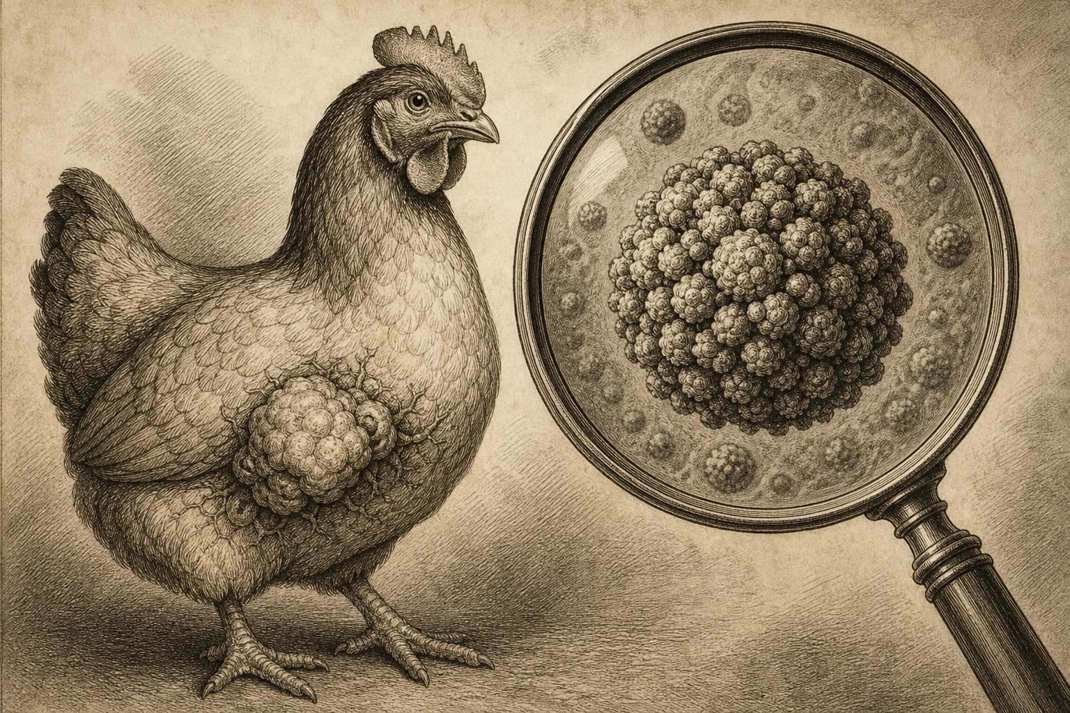
In 1976, Harold Varmus, J. Michael Bishop and Dominique Stehelin showed that a cancer-causing gene carried by a virus that gives chickens tumours was nearly identical to a gene in the normal cells of healthy birds. The virus had picked it up from its host. Cancer genes are faulty versions of our own.
A mutation can jam that accelerator down. Watch one cell keep dividing after its neighbours have stopped.
A virus from chickens Rous sarcoma virus carries the SRC gene; the same gene turned up in fish and mammals, including humans. AI-generated illustration.
SRC
the first oncogene found; its normal version is called a proto-oncogene
Nobel Prize, 1989
normal: growth on demand
mutated: growth signal stuck “on”
the virus copied it from a host cell
Proto-oncogene → oncogene
Chapter II · Accelerator and brakes
Oncogenes
In 1976 Varmus, Bishop and Stehelin reported in Nature that SRC, the oncogene of Rous sarcoma virus, “was nearly identical to a sequence in the normal cellular DNA” of several bird species: the virus had picked up a host gene. Bishop and Varmus shared the 1989 Nobel Prize “for their discovery of the cellular origin of retroviral oncogenes”.
The accelerator-and-brakes picture is a teaching analogy.
The 3D models are schematic and use illustrative numbers.
Other genes are the brakes, called tumour suppressors. We carry two copies of each, one from each parent, and one working copy is usually enough to stop the car. A “hit” here means a mutation that breaks one copy.
In 1971, Alfred Knudson studied a rare eye cancer in children. Inherited cases came younger, often in both eyes. His explanation: it takes two hits, one to each copy, and some children are born with the first hit already.
Watch twenty lives unfold. Those born with one broken copy reach their second hit much sooner.
the cell’s brakes
two copies, one from each parent
both must fail before control is lost
Tumour suppressor genes
inherited first hit present from birth; younger, often both eyes
sporadic both hits acquired during life; later onset
later the gene was identified as RB1
RetinoblastomaPone hit already≈pPnone yet≈p2
p
small chance that a given copy is hit
Why one hit makes it earlier (toy model)
Chapter II · Accelerator and brakes
Knudson’s two-hit hypothesis
Alfred G. Knudson proposed in 1971, from a statistical study of retinoblastoma, that two hits to DNA are needed. In inherited cases one hit is present from birth, so only one more is needed; sporadic cases need two acquired hits, which explains their later onset. The gene was later identified as RB1.
In the grid, the hit rate is invented for illustration; real retinoblastoma is rare.
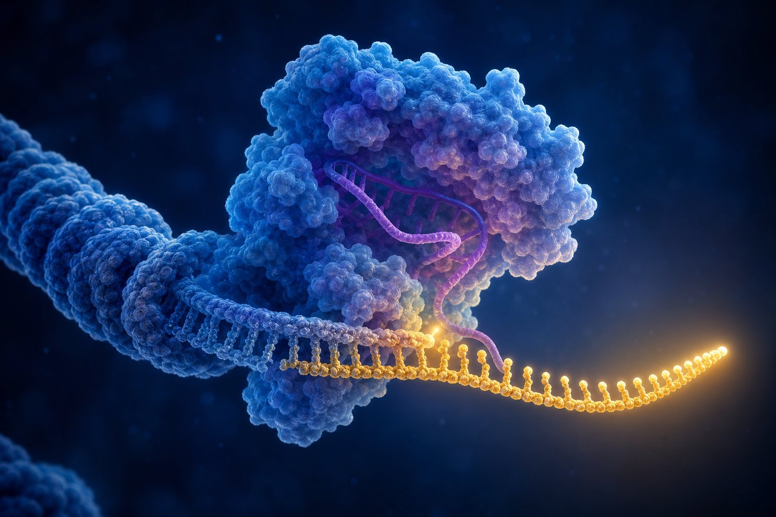
One brake matters more than most. A protein called p53, identified in 1979, watches for damaged DNA.
When it finds damage, it halts the cell until the damage is repaired, or, if the damage is too great, it tells the cell to destroy itself. The yellow cells here are damaged; watch them vanish or recover.
Now break p53. Damaged cells survive and divide, passing their mistakes on. It is no surprise that p53 is mutated in roughly half of all tumours.
senses DNA damage
pauses the cell cycle for repair
triggers apoptosis if repair fails
What p53 does
1979
first identified
~½
of tumours carry p53 mutations
p53
Chapter II · Accelerator and brakes
p53, guardian of the genome
p53 is a protein of 53 kilodaltons first identified in 1979. Mutations in p53 appear in roughly half of all tumours, and it is now known as the “guardian of the genome”.
The repair-or-remove choice in the model is simplified; real cells use many signals.
The 3D models are schematic and use illustrative numbers.
One stuck accelerator or one broken brake is rarely enough. A cell usually needs several hits in the right genes.
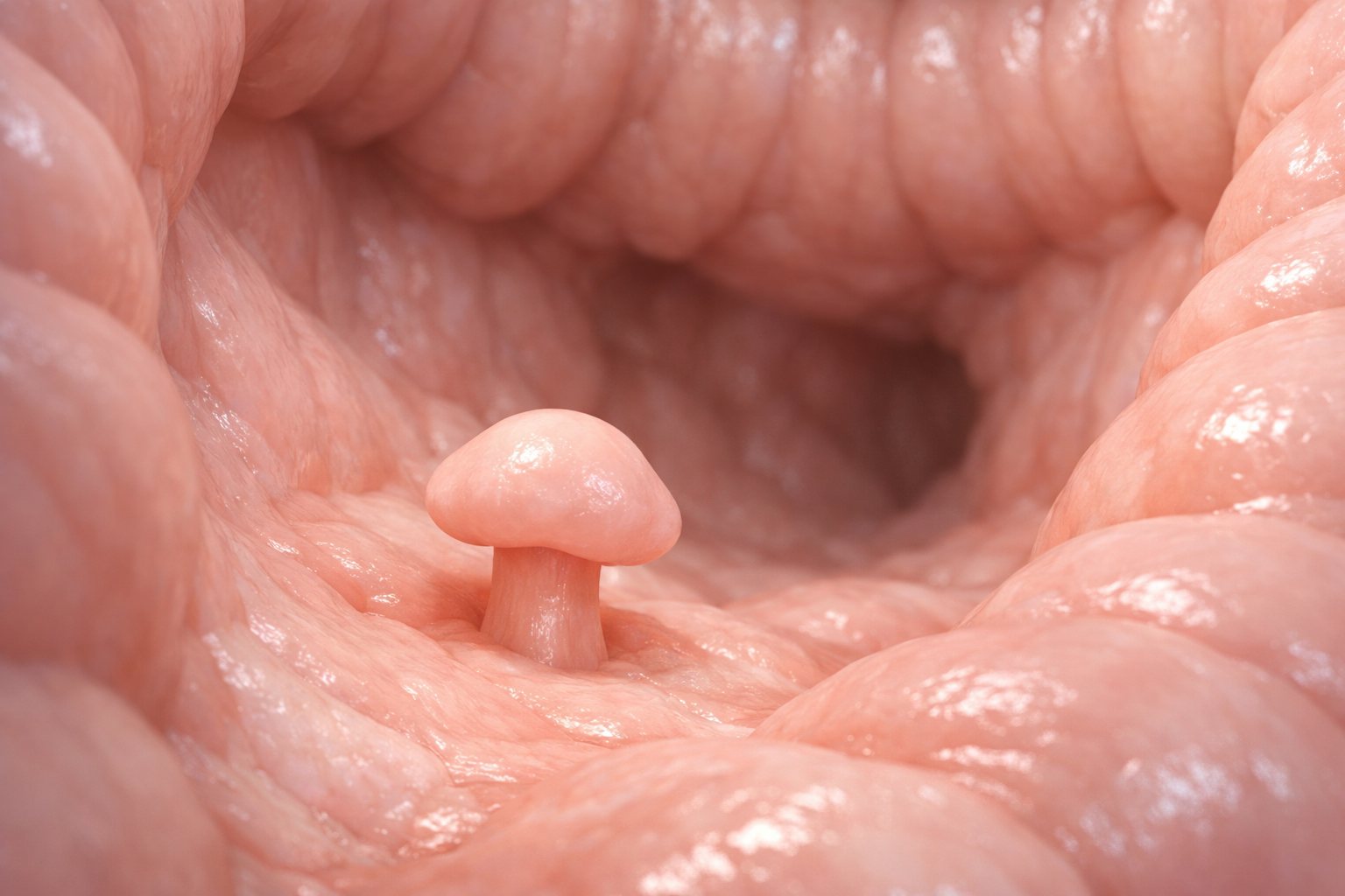
Bowel cancer shows the path most clearly. In a model proposed in 1990, a brake gene called APC fails first and a small growth called a polyp appears. Later, an accelerator gene, KRAS, and then p53 are hit.
Each step takes time. Studies estimate that going from a polyp to cancer typically takes more than a decade.
step 1 APC brake lost: a small polyp forms
step 2 KRAS accelerator jammed (in about 40% of tumours)
later p53 lost (over 60% of cancers): cancer
A model of bowel cancerA polyp Many polyps never become cancer, and they can be found and removed early. AI-generated illustration.
~10–26 years
range of estimated mean times
From polyp to cancer
Chapter III · Hit after hit
A stepwise disease
The Fearon–Vogelstein model (1990) holds that inactivation of the APC tumour suppressor occurs first in normal colonic tissue; activating KRAS mutations (found in about 40% of tumours) follow, and TP53 mutations (in over 60% of colorectal cancers) are late events. A review estimates a mean of 10.6 to 25.8 years from adenoma to cancer across all age groups.
Not every bowel cancer follows this exact order; it is the classic model.
Even a rogue cell faces another limit. In 1961, Leonard Hayflick found that normal human cells in a dish divide roughly forty to sixty times, then stop.
The clock is at the ends of each chromosome. Caps called telomeres lose a little length with every copy, and when they get too short, the cell retires.
Most cancer cells cheat by switching on an enzyme, telomerase, that rebuilds the caps. Switch it on, and the counter never runs out.
40–60
divisions before normal human cells stop, in culture
The Hayflick limit
repeated DNA at chromosome ends
a little is lost at every copy
too short → the cell becomes senescent
TelomeresRebuilt caps With telomerase active, the caps stay long, one of the hallmarks called replicative immortality. AI-generated illustration.
Chapter IV · The tumour’s tricks
Replicative immortality
Leonard Hayflick advanced the concept in 1961: normal human fetal cell populations divide roughly 40 to 60 times in culture before senescence. The limit correlates with telomere length, which shrinks with each replication. Most cancer cells do not senesce because they express telomerase, which extends telomeres.
The 3D models are schematic and use illustrative numbers.
A growing clump of cells soon runs short of food and oxygen. Without a blood supply it stays a tiny, pinhead-sized sphere.
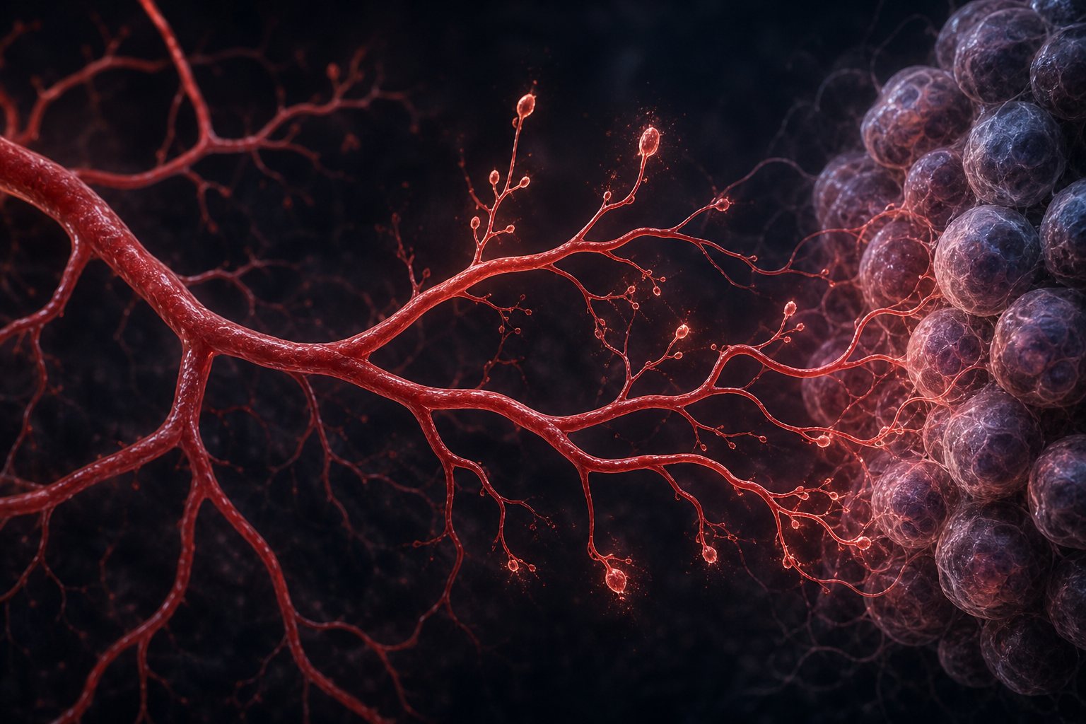
In 1971, the surgeon Judah Folkman proposed that tumours send out signals that make new blood vessels grow toward them. Watch the vessels arrive, and the tumour start to swell.
He also suggested that blocking those signals could keep tumours small, an idea that later led to new medicines.
Vessels sprouting Folkman called the signal “tumour angiogenesis factor”. AI-generated illustration.
the growth of new blood vessels
normal in healing and growth
hijacked by tumours
Angiogenesis
Chapter IV · The tumour’s tricks
Angiogenesis
In a 1971 paper in the New England Journal of Medicine, Judah Folkman proposed that tumours grow new blood vessels by releasing a signalling factor, and that blocking this process could keep tumours small. Without a blood supply, tumours stay as tiny, pinhead-size spheres.
“New medicines” refers to anti-angiogenic drugs used for some cancers; this film does not discuss treatment choices.
The 3D models are schematic and use illustrative numbers.
Not every tumour is cancer. A benign growth stays where it started. It can grow large and press on its neighbours, but it does not spread.
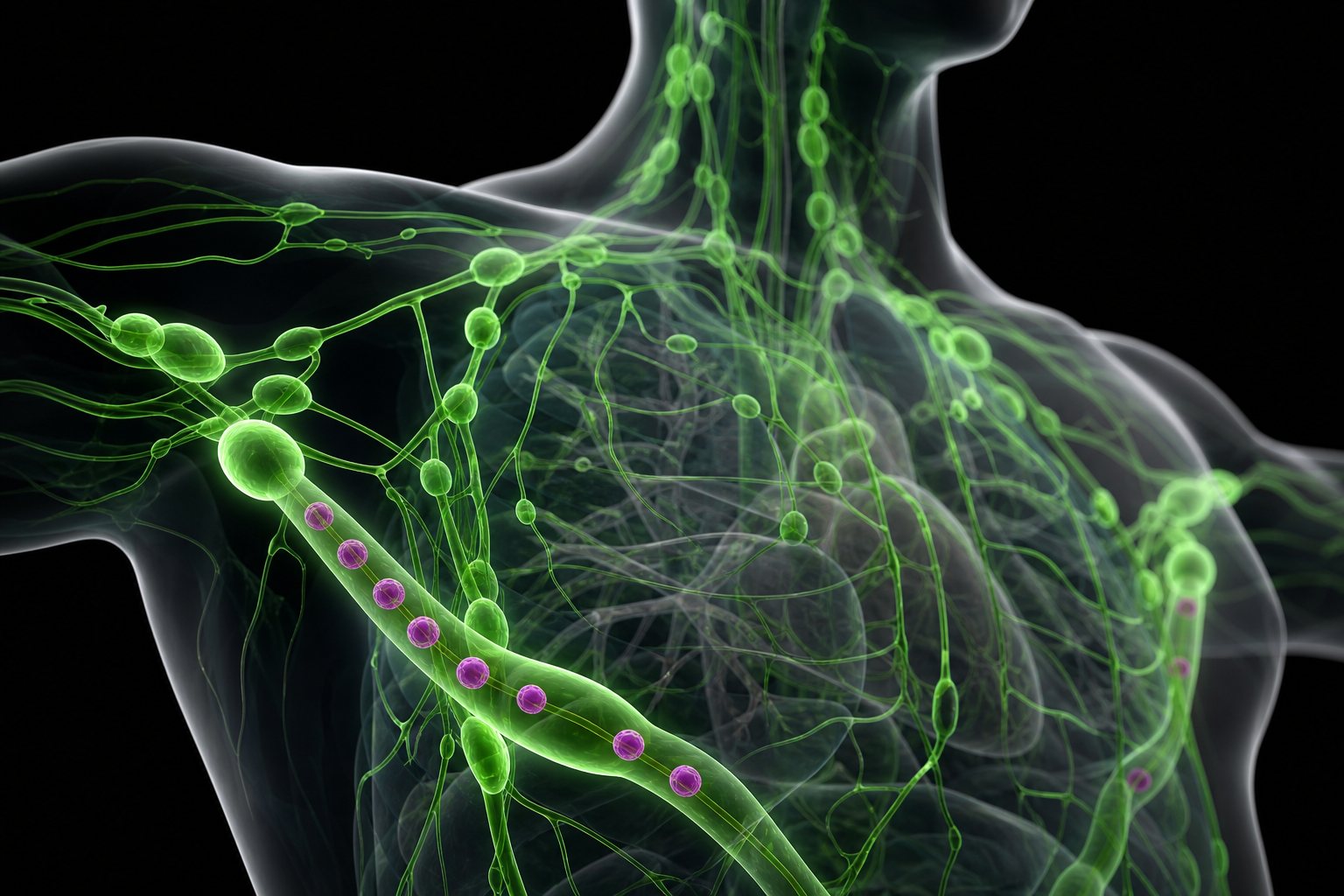
A malignant tumour invades the tissue around it. Its cells can slip into the bloodstream or the lymphatic system and settle in distant organs. This is metastasis.
Even far from home, the cells keep their origin. Breast cancer that has spread to the lungs still looks and behaves like breast cancer.
stays in its original place
often enclosed in a capsule
may press on nearby structures
BenignBlood and lymph To spread, a cell must leave the tumour, survive the journey and grow in a new tissue. AI-generated illustration.
100+
different diseases; each keeps the name of where it started, even after spreading
Cancer
Chapter IV · The tumour’s tricks
Benign and malignant
Benign growths stay in their original location; they may grow large and press on nearby structures but usually do not spread and are often enclosed in a capsule. Malignant tumours invade and destroy the tissue where they start, then spread and form new growths. Cancer keeps the characteristics of its organ of origin after it spreads. Cancer is more than 100 different diseases.
The 3D models are schematic and use illustrative numbers.
In 2000, Douglas Hanahan and Robert Weinberg gathered these tricks into a list they called the hallmarks of cancer: abilities that almost every cancer must acquire, whatever the organ.
Making its own go signals. Ignoring stop signals. Escaping self-destruction. Dividing without limit. Building blood vessels. Invading and spreading.
Later updates added rewiring the cell's energy use and hiding from the immune system.
self-sufficient growth signals
insensitive to anti-growth signals
evading apoptosis · limitless division
sustained angiogenesis
tissue invasion and metastasis
The six original hallmarks (2000)
2011 energy metabolism, immune evasion as emerging hallmarks; inflammation as enabling
2022 those two promoted to core hallmarks
Updates
Chapter IV · The tumour’s tricks
The hallmarks of cancer
Hanahan and Weinberg’s original six hallmarks (2000): self-sufficiency in growth signals, insensitivity to growth-suppressive signals, evading programmed cell death, replicative immortality, sustained angiogenesis, and tissue invasion and metastasis. In 2011 they added reprogramming energy metabolism and evading immune destruction as emerging hallmarks, and tumour-promoting inflammation as an enabling characteristic; in 2022 the two emerging hallmarks became core hallmarks.
Now we can answer the opening question. If a cell needs several rare hits, and hits arrive at a steady rate, then the chance that some cell has collected them all rises steeply with age.
Each dot is a family of cells descended from one ancestor. Move through the ages and watch the red dots, families with four hits, our stand-in for enough, stay rare for decades and then multiply.
That is what the statistics show. In the UK, more than half of new cancers are diagnosed in people aged seventy and over.
P(all k hits by age t)∝tk
k
number of hits needed
t
age
Needing k hits (rough idea)
> ½
of new cancers diagnosed at age 70+
85–89
age group where incidence peaks
UK, Cancer Research UK
Chapter V · Who gets it
Age and cancer
Cancer Research UK explains that DNA faults the body fails to repair accumulate over time, and each extra mutation makes a cell more likely to become cancerous. More than half of new UK cancer cases are diagnosed in people aged 70 and over, and incidence peaks at ages 85–89.
It also notes that mutations alone do not explain everything: ageing brings other changes, such as more inflammation. The dot model and the t^k rule are rough illustrations.
Worldwide, about twenty million people were diagnosed with cancer in 2022, and about nine point seven million died of it.
Many hits are bad luck. But in February 2026, the World Health Organization's cancer agency estimated that thirty-seven per cent of new cases were linked to preventable causes.
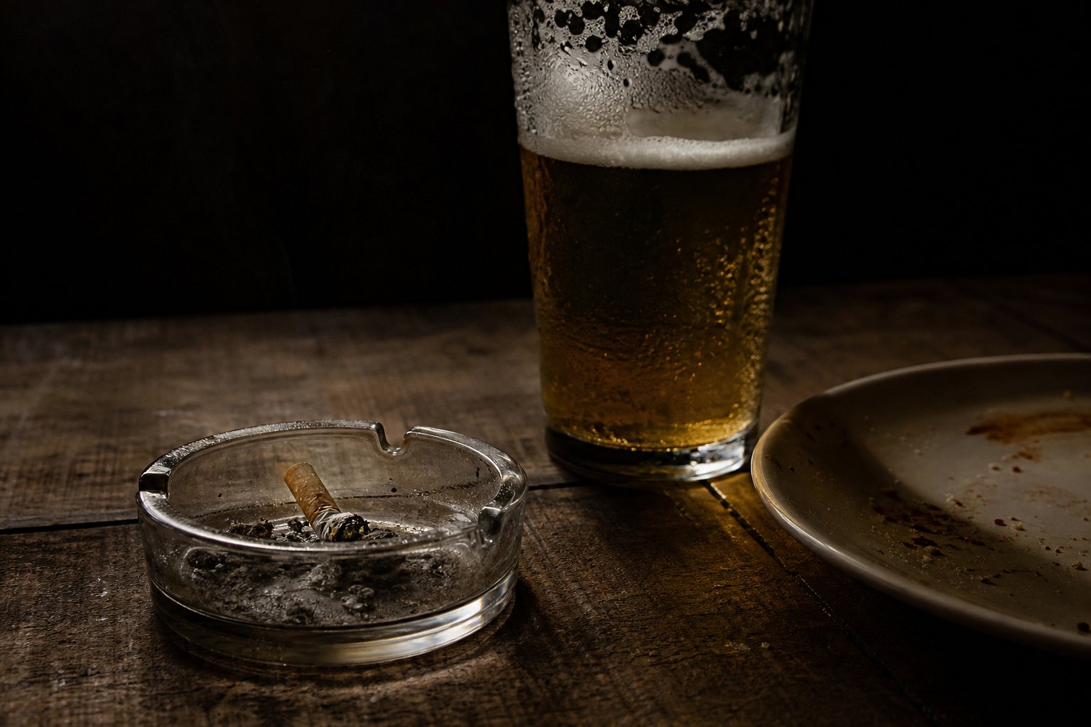
Tobacco led, linked to about fifteen per cent of all cases, then infections such as HPV and hepatitis at ten, and alcohol at three. Those are carcinogens raising the rate of hits.
~20 million
new cases
~9.7 million
deaths
GLOBOCAN 2022
all preventable causes
37%
tobacco
15%
infections
10%
alcohol
3%
Share of all new cases linked to…
Avoidable hits In men the WHO estimate rises to 45% of cases; in women it is 30%. AI-generated still.
Chapter V · Who gets it
The global picture
IARC’s GLOBOCAN estimates about 20 million new cancer cases and 9.7 million deaths in 2022. A WHO/IARC analysis released on 3 February 2026 linked 37% of new cases in 2022 (about 7.1 million) to 30 preventable causes, led by tobacco (15%), infections (10%) and alcohol (3%); 45% in men and 30% in women.
These are population estimates, not personal risk figures. For personal advice, talk to a doctor.
So what about younger adults? If more people are collecting hits earlier, cancers that usually wait until old age would start to appear sooner.
The analysis points to obesity, diet, inactivity, alcohol and smoking, and possibly the environment, while lung cancer in the young is falling as smoking declines. Some of the rise is better detection.
No one yet knows the full answer. A new British trial of a quarter of a million people aims to separate the causes.
kidney, 2000
2.5
kidney, 2023
5.3
US early-onset cancer, 2000 → 2023
obesity and diet
inactivity, alcohol, smoking
possibly environmental exposures
earlier detection (part of the rise)
Suspected contributorsFinding out A King’s College London trial funded by Cancer Research UK will follow 250,000 people, focusing on bowel cancer. AI-generated still.
Chapter V · Who gets it
Early-onset cancer
In the US, early-onset kidney cancer rose from 2.5 to 5.3 per 100,000 between 2000 and 2023, and bowel, uterine and pancreatic cancers each rose by more than 50%. Lung cancer in under-50s fell in all three countries. The researchers list obesity, diet, inactivity, alcohol, smoking and possibly environmental exposures as likely contributors; the analysis does not establish causes.
The first line of narration is a way of thinking about the trend, not a finding of the study.
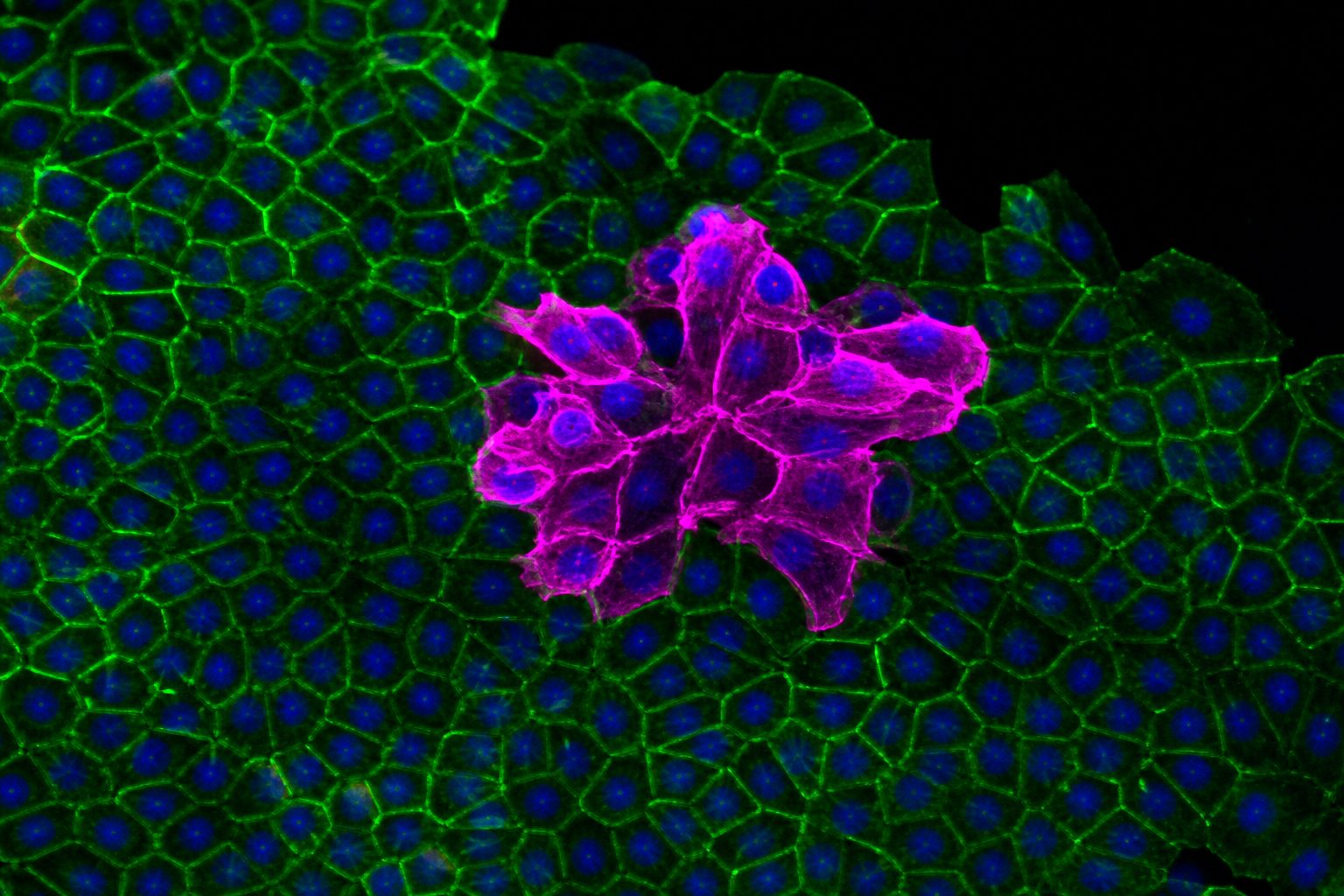
Cancer is not an invader. It is one of our own cells that, hit by hit, has jammed its accelerator and lost its brakes, its guardian and its countdown, then learned to feed itself and travel.
That takes many rare accidents, which is why it usually takes decades, and why the body stops almost every faulty cell long before.
Why some younger adults now seem to collect their hits sooner is still an open question. Each step in this film is a place where researchers are looking for the answer.
jammed an accelerator (oncogene)
broken both copies of a brake
escaped p53 and self-destruction
rebuilt its telomeres
grown vessels, invaded, spread
What a cancer cell has done
1961 Hayflick: normal cells divide 40–60 times
1971 Knudson’s two hits; Folkman’s angiogenesis
1976 SRC: cancer genes come from normal genes
1979 p53 identified
1990 stepwise model of bowel cancer
2000 the hallmarks of cancer
How we learnedOne cell out of step All stills and illustrations in this film are AI-generated.
Epilogue · Sources
How cancer starts
Cancer arises when a cell accumulates changes in genes that control growth, division, repair and death, and then acquires the abilities summarised as the hallmarks of cancer. Age, chance, inherited variants and preventable exposures all change how often those changes happen.
This film is general education, not medical advice, diagnosis or a guide to treatment. If you are worried about symptoms or your risk, talk to a doctor. All interactive models are schematic and all images are AI-generated.