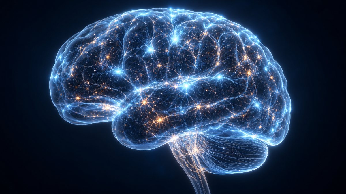
A new study links the APOE4 gene variant to a protein that weakens the brain's blood vessel barrier, in mice and human tissue. To see why that matters, this film explains Alzheimer's disease from the start: Alois Alzheimer's first patient in 1906, how neurons and synapses hold memories, how amyloid plaques and tau tangles form, how damage spreads through the brain over decades, why age and APOE4 raise the risk, and how the disease is diagnosed and treated today. Add amyloid and tau, step through the Braak stages, choose APOE versions and open the blood–brain barrier yourself. Education, not medical advice.
Interactive 3D film8 min1901 to 2026Published 20 October 2026
Some people carry a gene that makes Alzheimer's far more likely, yet stay sharp into their eighties. Why?
In 2026, a Columbia University team linked that gene, APOE e4, to a protein called fibronectin that weakens the wall between blood and brain.
First, though: what does Alzheimer's do to a brain?
~1 in 5
people carry at least one copy (Columbia team)
APOE e4
Bhattarai, Yilmaz, Kizil and colleagues, Columbia University; reported 19 Oct 2026
e4 mice: nearly twice the fibronectin of e3 mice
their blood–brain barrier leaked
mice, zebrafish, human tissue; no treatment tested
The new study (Nature Aging, 2026)Sharp at eighty Carrying e4 raises the odds; it does not decide them. AI-generated still.
Prologue · The news
APOE4, fibronectin and a leaky barrier
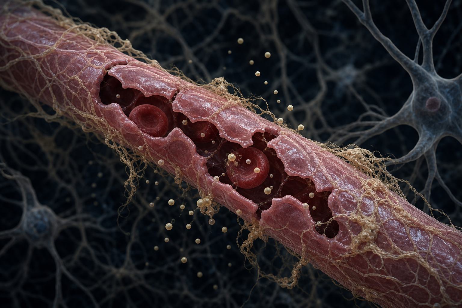
A study in Nature Aging by Prabesh Bhattarai, Elanur Yilmaz, Caghan Kizil and colleagues at Columbia University Irving Medical Center, reported by ScienceDaily on 19 October 2026 (ScienceAlert covered it on 18 September), found that APOE4 promotes fibronectin build-up around brain blood vessels. In mice carrying human APOE4, brain fibronectin was nearly double that in APOE3 mice and the blood–brain barrier appeared leaky; raising fibronectin in astrocytes made the barrier more permeable and reducing it improved barrier function.
This is one preclinical study: the mechanism comes from cells, zebrafish and mice, the human findings are correlational, and no treatment has been tested. This film is education, not medical advice; questions about your own health belong with a doctor.
How to use: pick a chapter or press ← →; drag to rotate. Nine steps have buttons you can press.
In November 1901, a fifty-one-year-old woman was admitted to an asylum in Frankfurt. Confused and forgetful, she told her doctor again and again: I have lost myself.
After she died in 1906, her doctor, Alois Alzheimer, examined her brain. Between the nerve cells were clumps, now called plaques. Inside the cells were twisted fibres, now called tangles.
Today, dementia means a decline in memory and thinking, and Alzheimer's is its most common cause, behind sixty to seventy per cent of cases.
Frankfurt, 1901 The patient was Auguste Deter, admitted on 25 November 1901. AI-generated still.
Nov 1901 Auguste Deter admitted, aged 51
Apr 1906 she dies, aged 55
Nov 1906 Alzheimer presents her case in Tübingen
1910 Emil Kraepelin names it “Alzheimer’s disease”
A disease gets a name
57 million+
people living with it
~10 million
new diagnoses a year
60–70%
of cases are Alzheimer’s
Dementia worldwide (WHO, 2026)
Chapter I · “I have lost myself”
Auguste Deter
Auguste Deter was admitted to the Frankfurt asylum on 25 November 1901, aged 51, and repeated “Ich habe mich verloren” (“I have lost myself”) during Alois Alzheimer’s examinations. She died on 8 April 1906. Examining her brain, Alzheimer found plaques and neurofibrillary tangles; he presented the case in Tübingen on 3 November 1906, and Emil Kraepelin named the disease in 1910.
WHO estimates that more than 57 million people live with dementia, with nearly 10 million new diagnoses a year, and that Alzheimer’s accounts for 60–70% of cases. Mayo Clinic calls Alzheimer’s the most common cause of dementia.
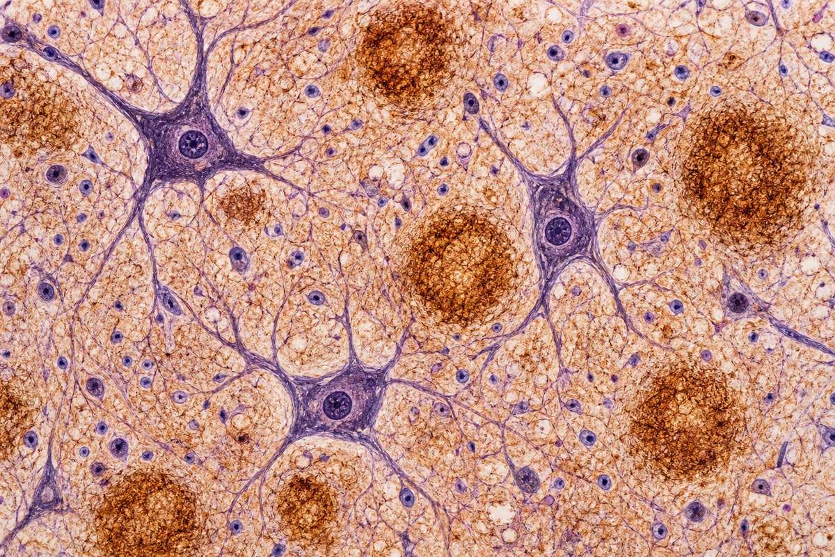
Start with the brain's wiring: about eighty-six billion nerve cells, or neurons, joined at roughly a hundred trillion contact points called synapses.
At a synapse, one neuron releases chemical messengers that tell the next whether to fire. Memories live in patterns of these connections.
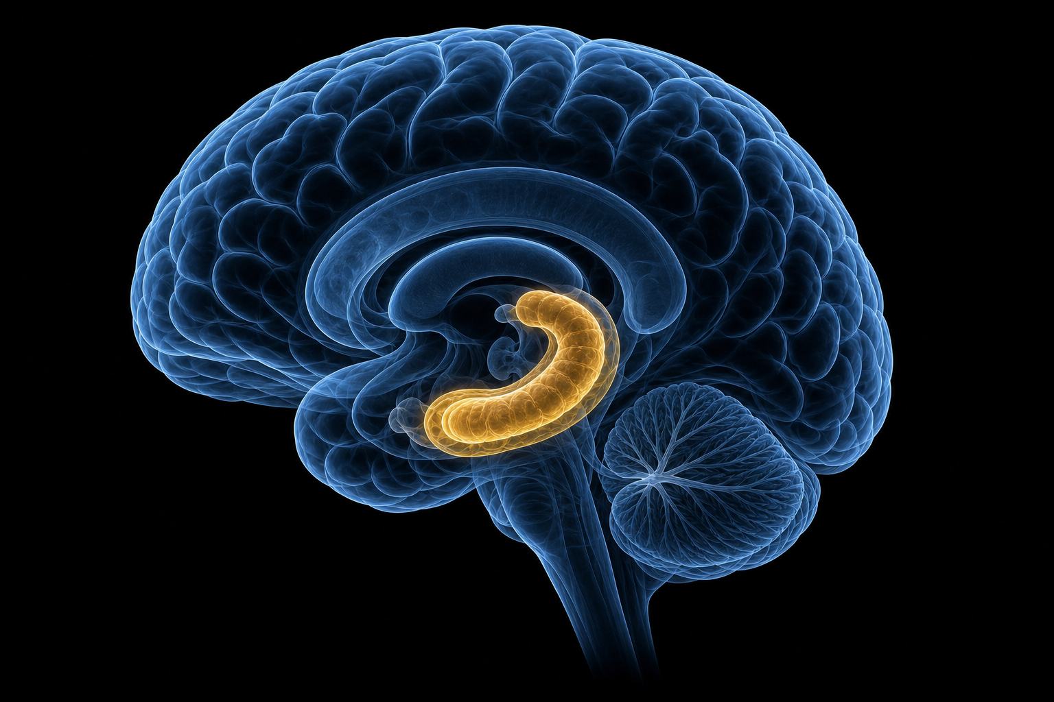
Deep in each side of the brain sits a seahorse-shaped structure, the hippocampus, which turns new experiences into lasting memories. Alzheimer's damage usually begins near it.
~86 billion
neurons
~100 trillion
synapses: thousands per neuron
The human brain
electrical: a pulse runs down the cell’s fibre
chemical: messengers cross a tiny gap
first mapped by Golgi and Cajal: Nobel Prize, 1906
One signal, two stepsThe hippocampus One in each temporal lobe; its name comes from the Greek for seahorse. AI-generated illustration.
Chapter II · The wiring of memory
Neurons, synapses and the hippocampus
The human brain has about 86 billion neurons and an estimated 100 trillion synapses, since each neuron makes thousands of connections. Golgi and Ramón y Cajal shared the 1906 Nobel Prize “in recognition of their work on the structure of the nervous system”. The hippocampus, deep in the medial temporal lobe of each hemisphere, helps form new memories and turns short-term memories into long-term ones.
Mayo Clinic notes that Alzheimer’s damage most often starts in the region of the brain that manages memory.
The plaques are made of a protein fragment, amyloid beta. Neurons carry a longer protein, APP, in their outer membrane, and enzymes snip pieces off it all the time.
Usually the cut is harmless. But when two other enzymes cut it in a different place, the leftover piece is sticky. It clumps into clusters, then fibres, then the plaques Alzheimer saw.
Add amyloid and watch clumps grow between the cells, as nearby signals begin to falter.
cut by α-secretase: harmless pieces that dissolve
cut by β-, then γ-secretase: amyloid beta, 40 or 42 building blocks (amino acids) long
Cutting APP (amyloid precursor protein)
1 single fragments (monomers)
2 small clusters (oligomers)
3 fibres (fibrils)
4 plaques between neurons
From fragment to plaqueAmyloid plaques George Glenner’s team characterised the plaque protein in 1984. AI-generated illustration.
Chapter III · Two proteins gone wrong
Amyloid beta
APP is a transmembrane protein. Cut by α-secretase it yields soluble fragments; cut first by β-secretase and then by γ-secretase it releases amyloid beta peptides of 40 or 42 amino acids, which are prone to form oligomers, then fibrils, then plaques. George Glenner’s team characterised the plaque protein in 1984.
Plaques are thought to impair synaptic transmission and trigger inflammation. McGill’s Brain from Top to Bottom notes that this “amyloid hypothesis” does not explain everything about the disease.
The second protein is tau. Inside each neuron, tracks called microtubules carry supplies out to its far ends, and tau holds those tracks steady.
In Alzheimer's, tau picks up too many chemical tags called phosphates. It is thought to let go of the tracks, which fall apart, while the loose tau knots into tangles.
Add tau and the deliveries stop. A neuron that cannot supply its synapses loses them, and may die.
binds the microtubule tracks
keeps transport running down the fibre
normal, healthy and essential
What tau does
1985
an antibody to tau was shown to mark the tangles (Jean-Pierre Brion)
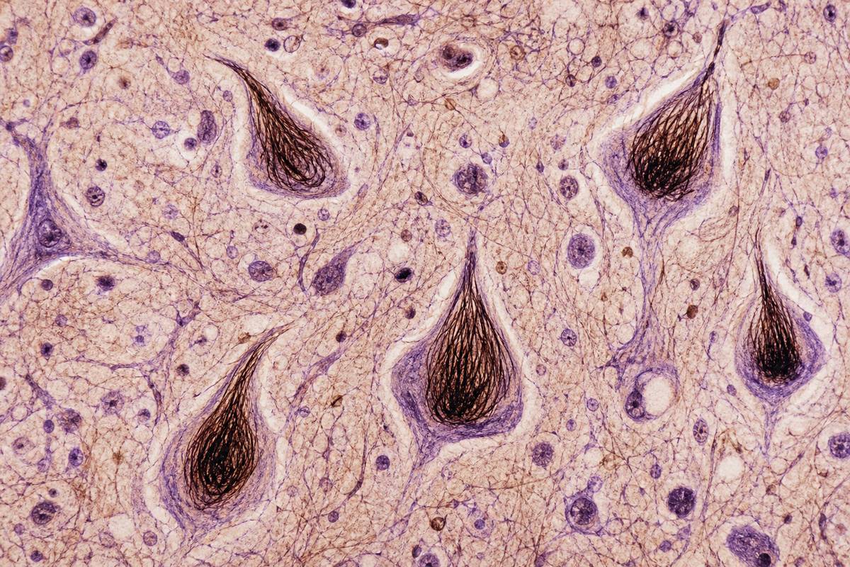
Tangles identifiedNeurofibrillary tangles Under a stain, tangles fill the cell body like dark flames. AI-generated illustration.
Chapter III · Two proteins gone wrong
Tau and tangles
Tau stabilises microtubules, supporting axonal transport and the integrity of the neuron. Neurofibrillary tangles consist of tau that has detached from microtubules and clumped together; excess phosphorylation is named as the main suspected cause. In 1985 Jean-Pierre Brion found an antibody that binds tau and marks the tangles, and other teams confirmed tau as their main component.
Tangles follow the same route in brain after brain.
Tangles do not appear everywhere at once. In 1991, two German anatomists, Heiko and Eva Braak, found that they follow the same route, in six stages.
In stages one and two, tangles sit beside the hippocampus; in three and four they reach it and the deep circuits of memory and emotion; in five and six, the cortex, the wrinkled outer layer.
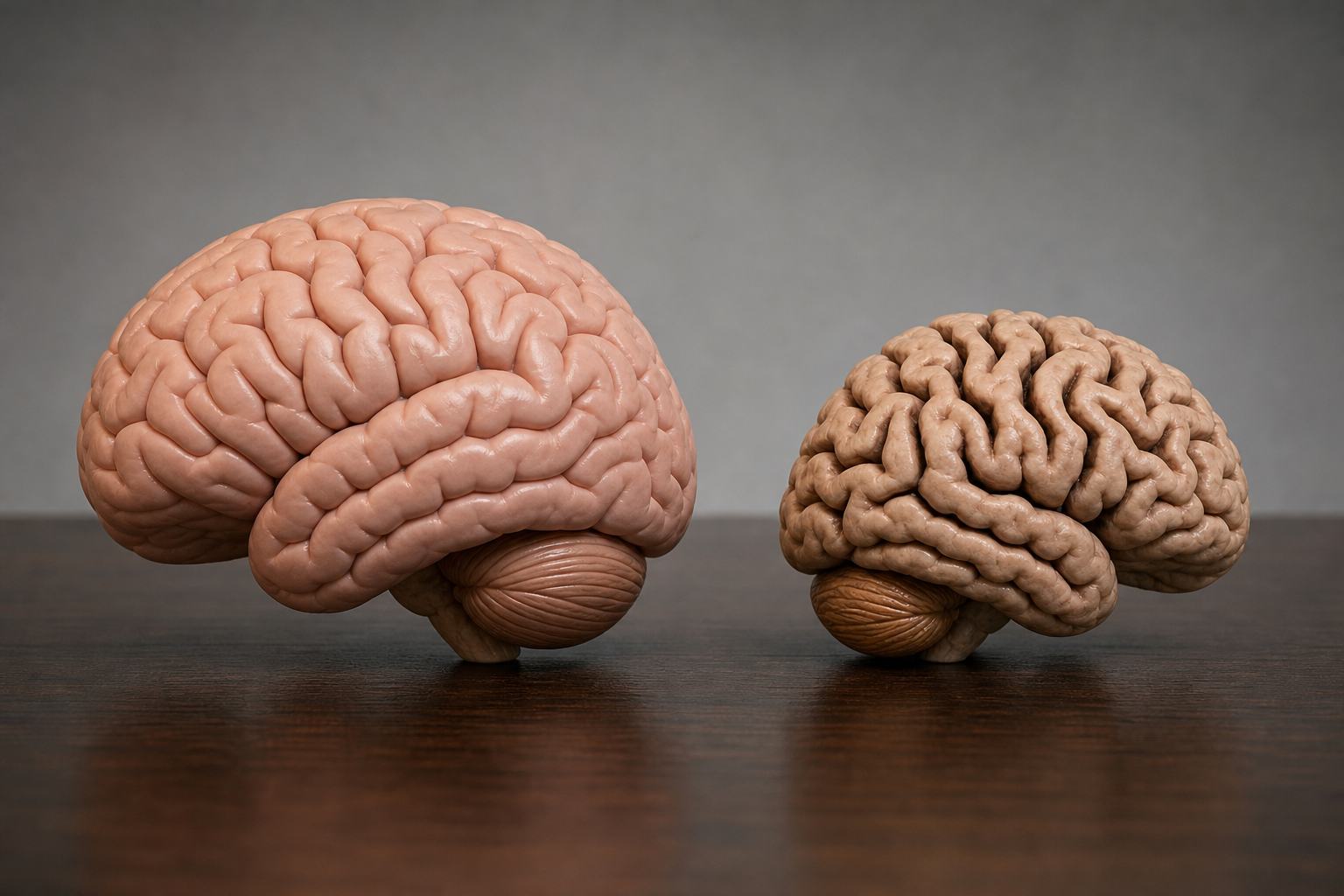
Step through the stages. As neurons die, the brain itself shrinks: late in the disease its folds are thinner and the grooves between them wider.
6
stages of tangle spread, seen after death
Braak & Braak, 1991
I–II a small area next to the hippocampus
III–IV the hippocampus and nearby memory and emotion circuits
V–VI most of the wrinkled outer cortex
The route of the tanglesA shrinking brain Models of a healthy brain and one in late Alzheimer’s: narrower folds, wider grooves. AI-generated still.
Chapter IV · A slow spread
Braak staging
Braak and Braak (1991) staged Alzheimer’s by neurofibrillary tangles: in stages I–II tangles are confined mainly to the transentorhinal region; in III–IV they also involve limbic regions such as the hippocampus; in V–VI they extensively involve the neocortex. Plaques spread in a different pattern. By the late stage of the disease, the brain has shrunk.
In the model the darkening is schematic: it shows where tangles are, in the order Braak described, on an illustrative brain.
Why some people and not others? Age is the strongest known risk: most cases begin at sixty-five or older.
Genes matter too. In 1993, researchers linked Alzheimer's to APOE, a gene whose protein carries cholesterol and other fats around the brain. It has three common versions, e2, e3 and e4, and we inherit one copy from each parent.
Compared with e3, the most common, one copy of e4 roughly doubles or triples the risk, and two copies make it eight to twelve times higher.
E2 lowers it. Yet many e4 carriers never get the disease.
older age: the strongest known risk factor
late-onset: 65 or older, the vast majority
rare inherited forms (APP, PSEN1, PSEN2): 1% of cases or less
Age and onset
20–30%
carry one or two copies
~2%
carry two copies
40–65%
of people diagnosed carry e4
APOE e4 (Alzheimer’s Association, US)
e3/e3 (two copies of e3)
1×
one e4
2–3×
two e4
8–12×
Risk compared with e3/e3 (Mayo Clinic)
e4 is a risk factor, not a direct cause
many people with e4 never develop the disease
many people without e4 do
Risk, not destiny (Mayo Clinic)
Chapter V · Who is at risk
APOE and other genes
Older age is the strongest known risk factor. In 1993 Allen Roses’s team at Duke reported that the APOE e4 allele is more frequent in late-onset Alzheimer’s and that risk rises with the number of copies. Mayo Clinic: at least one e4 “doubles or triples the risk”, two copies make someone “8 to 12 times more likely”; e2 “reduces the risk”; e3 appears to have no effect. Rare variants in APP, PSEN1 and PSEN2 cause early-onset familial disease, 1% of cases or less.
These are population figures. Genetic testing and what it means for a person is a conversation for a doctor or genetic counsellor. In the model, the e2 column has no single published figure: its height only says “lower”.
Another risk gene works in the brain's cleaning crew, its immune cells: a rare variant of TREM2, found in 2013, roughly triples the odds.
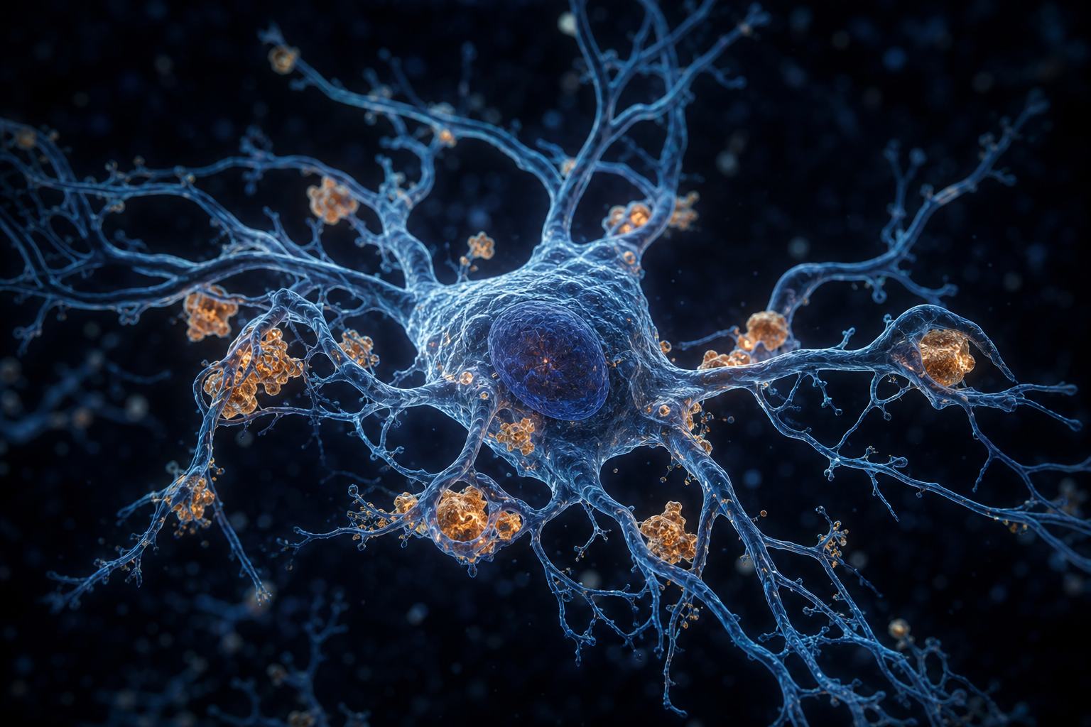
These cells, microglia, clear away debris. Early on they swallow amyloid and seem to help, but their inflammatory signals can also harm neurons.
Send in the microglia. Whether they mostly protect the brain or worsen the damage may depend on the stage of the disease.
2.9
odds ratio for the rare R47H variant (Icelandic study)
immune
a receptor on microglia
TREM2 (NEJM; online Nov 2012, print Jan 2013)A microglial cell Its branches constantly reach out, sampling the tissue around it. AI-generated illustration.
help: engulf amyloid and debris
harm: inflammation damages neurons
in one mouse study, removing them did not cut amyloid, yet fewer neurons were lost
Two faces of microglia
Chapter V · Who is at risk
Microglia and inflammation
Two studies in the New England Journal of Medicine (online 14 November 2012, in print January 2013) linked a rare TREM2 variant, R47H, to Alzheimer’s, with an odds ratio of about 2.9. In 2013 a variant of TREM2, a gene involved in inflammation and the immune response, was identified as a significant risk factor for late-onset Alzheimer’s (N Engl J Med 368:117–27). TREM2 is a receptor on microglia, the brain’s immune cells, which clean up debris and can consume plaques but also trigger inflammation that can damage nearby cells.
According to Scientific American, microglia appear protective in early stages and may become overwhelmed later; researchers are unsure whether they drive the disease or worsen a process amyloid starts.
Other things are linked to risk too. In 2024, the Lancet Commission on dementia estimated that up to forty-five per cent of dementia might be prevented or delayed by removing fourteen risk factors, from high blood pressure to hearing loss.
That is a population estimate, not a promise for anyone.
As countries age, this matters more: in Thailand, one person in five was sixty or older in 2024, about fourteen million people.
14
modifiable risk factors
up to 45%
of dementia potentially preventable or delayed
Lancet Commission, July 2024
less education · hearing loss · high LDL cholesterol · untreated vision loss
high blood pressure · diabetes · obesity · smoking
depression · inactivity · excess alcohol
head injury · air pollution · social isolation
The 14 factorsThailand, 2024 Share aged 60+: 6.8% in 1994, 20% in 2024 (NSO survey of older persons). AI-generated still.
Chapter V · Who is at risk
Modifiable risk
The 2024 Lancet Commission (published 31 July 2024) estimated that “45% of future dementia could potentially be prevented if all 14 risk factors were eliminated”: low education, hearing loss, hypertension, smoking, obesity, depression, physical inactivity, diabetes, excessive alcohol, traumatic brain injury, air pollution, social isolation, and two new ones, high LDL cholesterol and untreated vision loss. WHO guidelines in July 2026 likewise attribute up to 45% of dementia risk to modifiable factors.
These figures cover all dementia, not Alzheimer’s alone, and describe populations, not individuals. Nothing here is advice; for your own risk, talk to a doctor. Thai figures are from the National Statistical Office’s 2024 Survey of Older Persons, as presented by Chulalongkorn University’s Panthip Pruksacholavit.
The cruel part is timing. Two long studies published in 2024 followed people who later developed the disease.
Amyloid starts to build up first, roughly fifteen to nineteen years before symptoms. Tau changes follow about ten to fourteen years before. The hippocampus begins to shrink about seven years before diagnosis.
Only then do memory problems appear. Tap through the years with the buttons and watch each change arrive in turn.
Decades in silence One study followed 1,789 people for about 20 years. AI-generated illustration.
15–19 amyloid markers change (US study; 14–18 in China)
10–14 tau markers change
~7 hippocampus starts to shrink (before diagnosis)
~6 thinking tests start to decline
Years before symptoms or diagnosis (two 2024 studies)
US: Washington University, Annals of Neurology
China: COAST cohort, New England Journal of Medicine
amyloid → tau → shrinkage → symptoms
Same order in both studies
Chapter VI · Finding and treating it
Biomarkers before symptoms
Two studies published in February 2024, of sporadic Alzheimer’s, found the same order. Schindler, Li and colleagues (Annals of Neurology) estimated amyloid changes 19 to 15 years before symptoms and plasma p-tau217 14 to 12 years before; Jia and colleagues (NEJM; 1,789 people, about 20 years of follow-up) found amyloid changes 18 and 14 years, tau 11 to 10 years, hippocampal shrinkage 7 years and cognitive decline 6 years before diagnosis.
The model compresses this sequence into a few neurons; the timings on its clock follow the ranges above.
How is it found? Usually with a specialist's assessment and memory tests. But those measure symptoms, not proteins.
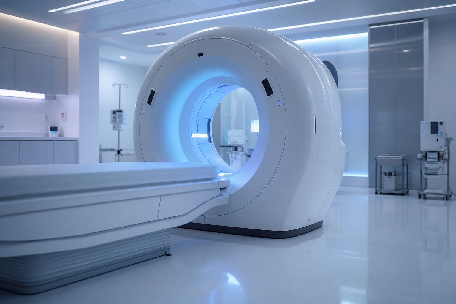
For a PET scan, a slightly radioactive tracer injected into a vein sticks to amyloid or tau and shows where it gathers. Spinal fluid can be tested too.
In May 2025, US regulators cleared the first blood test to help diagnose Alzheimer's, for adults fifty-five and over with symptoms.
history and a specialist’s assessment
memory and thinking tests
scans or fluid tests for the proteins
Steps towards a diagnosisA PET scanner Amyloid PET was the standard against which the new blood test was judged. AI-generated still.
91.7%
of positives had plaques on PET or spinal fluid
97.3%
of negatives had none
< 20%
results indeterminate
First FDA-cleared blood test, 16 May 2025 (study of 499 samples)A blood sample The cleared test compares two proteins in plasma: p-tau217 and amyloid beta 1-42. AI-generated still.
Chapter VI · Finding and treating it
Diagnosis
On 16 May 2025 the FDA cleared the Lumipulse G pTau217/β-Amyloid 1-42 Plasma Ratio for adults aged 55 and older with signs and symptoms of Alzheimer’s. In a study of 499 plasma samples from cognitively impaired adults, compared with amyloid PET or spinal fluid results, about 91.7% of positive results had amyloid plaques and 97.3% of negatives did not; fewer than 20% were indeterminate.
False positives and negatives occur, and the test shows whether plaques are present now, not what will happen. In the model, the amyloid and tau PET colours are schematic. Testing decisions belong with a doctor.
There is still no cure. Older medicines such as donepezil can ease symptoms for a time, but they do not stop the disease.
Newer drugs are antibodies that tag amyloid for clearing. Compared with a placebo, lecanemab slowed the worsening by about twenty-seven per cent over eighteen months; donanemab works similarly.
That is slower decline, not improvement. The benefit is modest, and both can cause brain swelling or small bleeds, called ARIA, most often with two copies of e4.
donepezil, galantamine, rivastigmine: boost a chemical messenger neurons use to signal (acetylcholine)
memantine: damps an excess of another messenger (glutamate)
ease some symptoms for a time; no cure
Older medicines (NHS)
Jul 2023 lecanemab: full US approval (decline 27% slower than placebo over 18 months)
Jul 2024 donanemab: US approval
2025 EU approves both, not for people with two e4 copies
Anti-amyloid antibodiesGiven by infusion Treatment needs confirmed amyloid and regular MRI scans to watch for ARIA. AI-generated still.
0.7%
of participants had severe ARIA
MRI
scans before and during treatment
ARIA (Clarity AD trial)
Chapter VI · Finding and treating it
Treatments
The NHS lists acetylcholinesterase inhibitors (donepezil, galantamine, rivastigmine) and memantine, which can temporarily reduce some symptoms; there is no cure. Lecanemab received full FDA approval on 6 July 2023 after the Clarity AD trial showed 27% less clinical decline than placebo at 18 months; donanemab was approved on 2 July 2024. The EU approved lecanemab in April 2025 and donanemab on 25 September 2025, for people with confirmed amyloid who carry no more than one e4 copy.
ARIA (amyloid-related imaging abnormalities) means temporary brain swelling or small bleeds seen on MRI; it is usually without symptoms but can be serious, and the risk is higher with two e4 copies. NICE, the cost watchdog for England and Wales, did not recommend either drug for routine NHS use in June 2025, judging the benefit too modest for the cost. After appeals it consulted on a third draft from 31 March to 28 April 2026, and its committee met again on 8 July 2026; at the end of July no new decision had been published, no deadline was set, and price talks with NHS England were under way. Whether a treatment is suitable is a decision for a doctor.
Back to the news. The brain's smallest blood vessels are sealed by their lining and wrapped by support cells called astrocytes. This blood–brain barrier keeps most blood proteins and immune cells out.
When it leaks, they get in and can fuel inflammation that harms neurons. In mice with the human e4 gene, the Columbia team found nearly twice the fibronectin seen with e3, and a leaky barrier. Try it: switch to e4, then reduce fibronectin.
That may be part of the answer to our first question: some healthy e4 carriers have a rare protective change in the fibronectin gene. But this is one study in animals and tissue, and fibronectin also helps wounds heal.
lining cells joined by tight junctions
astrocyte “end-feet” wrap the vessel
when it leaks: blood proteins such as fibrinogen and immune cells get in
The blood–brain barrierA leaky barrier In the study, excess fibronectin upset the growth signals that keep the barrier sealed. AI-generated illustration.
~2×
brain fibronectin in e4 mice compared with e3 mice
Mice with human APOE (Columbia study)
shown: a mechanism in cells, zebrafish and mice
human tissue: an association, not proof
not shown: any benefit for memory
no treatment tested
What the study does and does not show
Reducing fibronectin itself rescues the barrier defects.Caghan Kizil, Columbia University, to ScienceAlert (in animal models)
Chapter VII · Back to the news
Fibronectin and the barrier
The blood–brain barrier is made of endothelial cells joined by tight junctions, supported by a basement membrane, pericytes and astrocyte end-feet; it limits what passes from the blood into the brain. A 2023 review (Chen et al.) describes how, when it is disrupted in Alzheimer’s, plasma proteins such as fibrinogen and immune cells enter, inflammation increases, and barrier damage and disease may drive each other; APOE4 carriers show more barrier dysfunction. Kizil’s team found that APOE4 promotes fibronectin around brain vessels; excess fibronectin activated abnormal integrin and focal adhesion kinase signalling and disrupted VEGF, HBEGF and IGF1 signals that maintain the barrier.
The team’s earlier work found a rare protective FN1 variant in some APOE4 carriers. Fibronectin has essential roles in tissue support and healing, so any future treatment would have to target only its harmful build-up. The model is schematic; “nearly twice” is the mouse measurement.
Alzheimer could only look at his patient's brain after she died. Today the same proteins show up in a living brain, and in blood, years before the first forgotten name.
That silent stretch is where research now looks: earlier treatment, the brain's immune cells, and its blood vessels.
And for the sharp eighty-year-olds we began with, part of the answer may lie in those vessels. Researchers are now exploring that idea in the lab.
1906 Alzheimer describes plaques and tangles
1984–85 amyloid beta and tau identified
1991 Braak stages of tangle spread
1993 APOE e4 linked to risk
2023–25 anti-amyloid drugs; first blood test
2026 fibronectin and the barrier (mice)
How we learned
why some e4 carriers stay healthy
how amyloid, tau and inflammation drive each other
whether treating earlier helps more
memory changes? talk to a doctor; sources are in the panel
Open questionsA brain at work All stills and illustrations in this film are AI-generated.
Epilogue · Sources
How Alzheimer’s disease works
Alzheimer’s disease is defined by amyloid plaques between neurons and tau tangles inside them. The changes begin one to two decades before symptoms, spread through the brain in a regular order, and end in the loss of synapses and neurons. Age is the strongest risk factor; APOE e4 raises risk; diagnosis can now detect the proteins in life, and anti-amyloid antibodies modestly slow early disease.
This film is general education, not medical advice, diagnosis or a guide to treatment. If you are worried about memory changes, talk to a doctor. All interactive models are schematic and all images are AI-generated.