1952 → 2026 · Nobel Prize in Physiology or Medicine
Switching On the Brain
How a neuron fires, how a green pond alga senses light, and how its light-gated channel became optogenetics, the tool that switches chosen brain cells on and off with light. The story behind the 2026 Nobel Prize in Physiology or Medicine.
Interactive 3D film7 min1952 to 2026Published 2026-10-13
October 2026 · Nobel Prize in Physiology or Medicine
Switching on the brain
Three scientists found a way to turn chosen brain cells on and off with light.
On the fifth of October 2026, the Nobel Prize in Physiology or Medicine went to Karl Deisseroth, Peter Hegemann and Georg Nagel.
It honoured their discoveries on light-gated ion channels and optogenetics: a way to switch chosen brain cells on and off with light.
Your brain holds about eighty-six billion neurons, signalling to one another in tiny electrical pulses.
To see how a flash of light can control them, we start with how a single neuron sends a signal.
Prologue · Why the news matters
Switching on the brain
On 5 October 2026 the Nobel Assembly at Karolinska Institutet awarded the Nobel Prize in Physiology or Medicine to Karl Deisseroth (Stanford University), Peter Hegemann (Humboldt University of Berlin) and Georg Nagel (University of Würzburg) “for their discoveries concerning light-gated ion channels and optogenetics.” Their work lets scientists switch the activity of individual kinds of nerve cells on or off with light, with millisecond timing.
Neurons in a human brain
~86 billion
plus a similar number of non-neuronal cells
Prize
12 million SEK
shared by the three laureates
The idea in one line
Borrow a light-sensitive channel from a green alga, put its gene into chosen neurons, and flash light to make them fire.
The brain on the left is drawn as a cloud of points; each point stands for millions of real neurons. Flickers show cells firing.
How to use: pick a chapter below the scene or press ← → to change step · ▶ plays the step · drag to rotate, scroll or pinch to zoom · buttons in the box at the top right let you run the experiments yourself · “Play the film” runs every step full-screen.
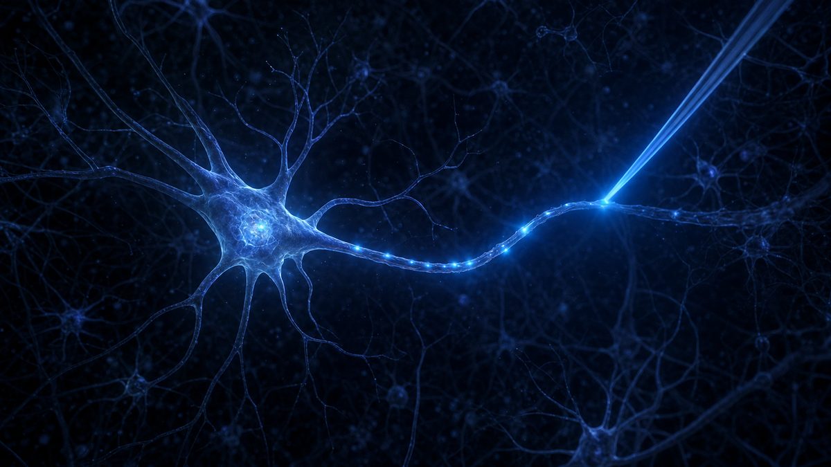
A neuron gathers signals with its branches and sends its answer down one long cable.
A neuron is a cell built for messages. Its branching dendrites gather signals from thousands of other cells.
If the input is strong enough, the cell fires. A pulse races down its long axon toward the next cells.
Where the axon ends, at synapses, it releases chemical messengers that excite or calm its neighbours.
The pulse itself is electrical. But it is not carried by electrons in a wire. It is carried by ions.
Chapter I · Step 1
A living wire
A neuron has three working parts: branching dendrites that receive signals, a cell body that adds them up, and a long axon that carries the cell’s own signal away. At the axon’s endings, synapses pass the message to the next cells with chemical messengers called neurotransmitters.
Inputs arrive at the dendrites and nudge the cell’s voltage up or down.
If the voltage at the cell body reaches a threshold, the neuron fires an all-or-nothing pulse: an action potential.
The pulse runs along the axon. A fatty sheath called myelin lets it jump from gap to gap, which is faster and saves energy.
At synapses, glutamate usually excites the next cell; GABA usually calms it.
The trace at the top left is the cell body’s voltage, slowed down so you can follow it. Each tall spike is one action potential.
Every neuron keeps its inside slightly negative, like a tiny battery waiting to discharge.
The neuron is wrapped in a thin, oily membrane. Charged atoms, called ions, cannot slip through it on their own.
Protein pumps spend energy pushing three sodium ions out for every two potassium ions they bring in.
So sodium piles up outside and potassium inside, and some potassium slowly leaks back out through open channels.
The result is a voltage: the inside sits about seventy millivolts below the outside. The cell is charged and ready.
Chapter I · Step 2
A charged skin
The cell membrane is a double layer of fatty molecules. Ions carry electric charge, so they cannot cross this oily barrier by themselves. They pass only through proteins: pumps that use energy to move them, and channels, pores that open and close.
Na⁺ sodiumMostly outside the cell
K⁺ potassiumMostly inside the cell
Cl⁻ chlorideMostly outside the cell
Sodium–potassium pump
3 out : 2 in
uses ATP, the cell’s energy currency
Resting voltage
about −70 mV
inside compared with outside
Press “Run the pump” to watch one cycle. Ions are drawn far larger and fewer than in reality.
In about a millisecond, gates in the membrane snap open and shut, and the voltage swings by a hundred millivolts.
Push the voltage up to a threshold, around minus fifty-five millivolts, and voltage-gated sodium channels snap open.
Sodium floods in. In well under a millisecond, the inside swings to about plus forty millivolts.
Then potassium channels open, potassium rushes out, and the voltage falls back. This all-or-nothing pulse is the action potential.
In 1952, Alan Hodgkin and Andrew Huxley worked this out with the giant nerve fibre of a squid. It earned them a Nobel Prize in 1963.
Chapter I · Step 3 · 1952
The spike
An action potential is a brief reversal of the membrane voltage. It is all or nothing: below threshold, nothing happens; above it, the cell always fires a full-sized spike.
Recovery: the voltage briefly dips below rest, then the pumps restore the balance.
Alan Hodgkin and Andrew Huxley measured these currents in the giant axon of the squid and described them with equations published in 1952. They shared the 1963 Nobel Prize with John Eccles “for their discoveries concerning the ionic mechanisms involved in excitation and inhibition” of nerve cells. Press “Stimulate” to fire a spike yourself.
To learn what one kind of cell does, you need to switch on that kind, and only that kind.
For a long time, the main way to make neurons fire on command was to pass electric current into the brain through a metal electrode.
But current does not choose. It excites every cell near the tip, along with fibres just passing through.
Drugs can be more selective, but they act over minutes, far too slowly to follow events that unfold in milliseconds.
In 1979, Francis Crick wrote that a real challenge for neuroscience was to control all cells of one type, while leaving the others more or less unaltered.
Chapter II · Step 4
Too blunt a tool
The brain packs many kinds of neurons side by side, wired into circuits. To find out what one kind does, you would like to switch on just those cells, at exactly the right moment, and watch what changes.
Electrode
Fast but unselective
excites every cell and fibre near its tip
Drugs
Selective but slow
take minutes to act and wear off
What was missing
A switch that is both fast (milliseconds) and specific (one cell type)
Francis Crick suggested in 1979 that light might one day provide the precision needed
In the scene, the electrode lights up every point near its tip, whatever its type. Press “Current on / off” to try it.
The tiny alga Chlamydomonas swims toward light. How does a cell with no nerves see?
The answer came from an unlikely place: a pond alga called Chlamydomonas, a single green cell that swims with two flagella.
It steers toward light using a small orange eyespot. Peter Hegemann set out to find the light sensor behind it.
Like our own eyes, it relies on rhodopsins: proteins that hold a light-catching molecule called retinal, made from vitamin A.
When light hits, retinal changes shape almost instantly, and the protein around it reacts.
Chapter III · Step 5
An eye in a single cell
Chlamydomonas reinhardtii is a single-celled green alga about a hundredth of a millimetre across. It swims with two flagella in a breaststroke and steers toward light that is good for photosynthesis. A patch of orange pigment, the eyespot, shades the light-sensing membrane from one side, so as the cell rolls it can tell where the light is coming from.
Peter Hegemann studied how this alga senses light, beginning in the early 1990s at a Max Planck institute.
Its photoreceptors are rhodopsins: proteins holding retinal, a molecule related to vitamin A, also used in animal eyes.
Absorbing light flips retinal from one shape to another (from all-trans to 13-cis), which changes the protein.
The scene shows the eyespot glowing when it faces the light. Sizes and speeds are not to scale.
The alga’s light sensor turned out to be a pore in the membrane, opened directly by light.
Working with Ernst Bamberg in Frankfurt, Georg Nagel put the alga’s genes into frog egg cells to test what the proteins did.
In 2002, with Hegemann, they showed that channelrhodopsin-1 is an ion channel opened directly by light. In 2003 came channelrhodopsin-2.
Shine blue light on it, around 460 nanometres, and its pore opens within a fraction of a millisecond. Positive ions, such as sodium, pour in.
No other parts are needed. Light in, current flows. It worked in frog eggs, and in human cells grown in a dish.
Chapter III · Step 6 · 2002–2003
A channel that opens with light
Georg Nagel had shown with Ernst Bamberg at the Max Planck Institute of Biophysics that a microbial rhodopsin could work in frog egg cells. Together with Hegemann they tested the alga’s rhodopsin genes. Channelrhodopsin-1 (2002) and channelrhodopsin-2 (2003) proved to be something new: ion channels gated directly by light.
Opened by
blue light
peak response near 460 nm
Opening time
< 0.2 ms
measured rise time about 200 µs or less
Lets through
positive ions
such as Na⁺, K⁺ and Ca²⁺, but not negative ions
Parts needed
one
a single protein; cells already contain retinal or can be given it
Because one gene makes the whole switch, it can be moved into almost any cell. Press “Blue light” to open the channel and watch the voltage rise.
The algal gene was put into rat neurons. Each blue flash made them fire, spike for spike.
At Stanford, Karl Deisseroth, a psychiatrist and bioengineer, wanted a way to control chosen brain cells directly.
With Ed Boyden, Feng Zhang, Nagel and Bamberg, his team used a harmless virus to carry the channelrhodopsin-2 gene into rat neurons.
Each flash of blue light made the neuron fire a spike, with millisecond timing. Their paper appeared in August 2005.
This is optogenetics: genes that make cells sensitive to light, plus optics that deliver the light exactly when you want.
Chapter IV · Step 7 · 2005
Light becomes a switch
In August 2005, Edward Boyden, Feng Zhang, Ernst Bamberg, Georg Nagel and Karl Deisseroth reported in Nature Neuroscience that neurons carrying channelrhodopsin-2, delivered with a lentivirus, could be made to fire by brief pulses of blue light, with millisecond precision. Light could now control both excitatory and inhibitory signals between cells.
Gene: the instructions for channelrhodopsin-2.
Delivery: a disabled virus carries the gene into neurons, which then build the channel in their membranes.
Light: a blue flash opens the channels, positive ions flow in, and the neuron reaches threshold.
Others were racing too. In 2002 Gero Miesenböck’s team had made neurons respond to light with three fruit-fly genes. Channelrhodopsin did it with one, and it was fast. Try “Blue flash” and “Pulse train” to fire the neuron.
A second protein from salt-loving microbes silences neurons with yellow light.
An on switch is only half a tool. Scientists also needed a way to switch neurons off.
They found it in halorhodopsin, from salt-loving microbes. Yellow light makes it pump chloride ions into the cell.
The extra negative charge pulls the voltage further down, so the neuron cannot reach threshold and stays silent.
By 2007, labs could put both proteins into the same cells: blue to switch them on, yellow to switch them off.
Chapter IV · Step 8 · 2007
An off switch too
Halorhodopsin comes from Natronomonas pharaonis, a microbe that lives in very salty lakes. It is a light-driven pump that moves chloride ions (Cl⁻) into the cell. In 2007 the Boyden and Deisseroth labs each showed that it can silence neurons.
On switch
ChR2
blue light (~470 nm) · lets positive ions in
Off switch
NpHR
yellow light (~560 nm in the 2007 study) · pumps Cl⁻ in
Turn “Yellow light” on, then press “Blue flash”: the blue flash still nudges the voltage up, but no longer enough to reach threshold. Two colours, two directions, in the same cell.
A genetic switch decides which cells make the light-sensitive protein. A fibre decides where the light goes.
In a real experiment, the gene for the light-sensitive channel is paired with a promoter: a stretch of DNA that is active only in one type of cell.
A virus delivers it into one small region of the brain. Only the chosen cell type builds the channel.
Then a hair-thin optical fibre brings light into that region, while the animal moves freely.
When the light comes on, only the tagged cells respond. Their neighbours, a few thousandths of a millimetre away, carry on as before.
Chapter V · Step 9
Only the cells you choose
Optogenetics gets its precision from two places at once: genetics chooses which cells can respond, and optics chooses where and when the light arrives.
Promoter: a DNA switch that is turned on only in one cell type, for example cells that make a particular neurotransmitter.
Virus: injected into one brain region, it carries the gene into cells there.
Optical fibre: implanted above the region, it carries laser or LED light. Fibre-coupled light sources for freely moving animals came into use in 2007.
Result: light switches the chosen cells on or off within milliseconds; other cells nearby are untouched.
Most of this research is done in mice. In the scene, tagged cells are shown in teal. Press “Light on / off” to compare the light with the electrode in Chapter II.
Laboratories around the world use optogenetics to find out which cells cause which behaviour.
Since then, optogenetics has spread to laboratories all over the world, working mostly with mice, flies, worms and fish.
Before, scientists could mainly watch which cells were active during a behaviour. Now they could switch those cells on or off, and see what changed.
That turned correlation into cause. It has been used to probe circuits for memory, fear, reward, movement, sleep and hunger.
It is also helping researchers study how circuits change in conditions such as Parkinson’s disease, addiction and depression.
Chapter V · Step 10
Reading the wiring
Recording tells you which cells are active when something happens. Optogenetics lets you test whether those cells cause it: switch them on and see if the behaviour appears, switch them off and see if it disappears.
Memory activating or silencing cells in the hippocampus to test how memories are stored and recalled
Emotion circuits through the amygdala involved in fear and anxiety
Reward dopamine cells deep in the midbrain and the motivation they drive
Movement circuits of the motor cortex and basal ganglia, important for understanding Parkinson’s disease
The highlighted regions in the scene are approximate and drawn on a simplified brain; most of these experiments were done in mice, not people. Press “Next circuit” to move the fibre.
The first report of partial vision returning to a person through optogenetic therapy.
Most uses of optogenetics are in research. But in the eye, light can reach the cells without surgery to place a fibre.
In retinitis pigmentosa, the eye’s light-sensing cells die. Other retinal cells, the ganglion cells, survive.
In a study published in 2021, a blind man received a gene for ChrimsonR, an amber-sensitive channelrhodopsin, in one eye.
Wearing goggles that turned the scene into amber light, he could find and touch a large notebook on a table in 36 of 39 tries.
Chapter VI · Step 11 · 2021
Light for a blind eye
In May 2021 a team from Paris, Pittsburgh and Basel reported in Nature Medicine the partial recovery of visual function in a man blinded by retinitis pigmentosa, an inherited disease that destroys the eye’s photoreceptors. A gene therapy made his surviving retinal ganglion cells produce ChrimsonR. Goggles with a camera projected the scene onto his retina as amber light, which is gentler on the retina than blue.
Find and touch a notebook
36 / 39
92% of trials, with goggles
A small staple box
36%
of trials
Count glass tumblers
63%
correct
Brain-signal check
78%
an EEG decoder could tell when a tumbler was present
This was one patient in an early-stage trial. It showed that the idea can work in a human, not that it is a cure. The retina in the scene is simplified.
A question about how algae swim toward light became one of the most powerful tools in biology.
Optogenetics grew out of curiosity about how a pond alga finds the light. Nobody set out to build a brain tool.
Many people shaped it, among them Ernst Bamberg, Ed Boyden, Feng Zhang, and Gero Miesenböck, who first made neurons respond to light in 2002.
A Nobel Prize can be shared by at most three people. This year it honoured the light-gated channel, and the method built on it.
Today, a flash of light can ask one kind of brain cell a simple question: what do you do? And the brain answers.
Epilogue
From pond alga to brain
The road to optogenetics ran through basic science that had no medical goal: how a single-celled alga senses light, and how ions cross a membrane. Combined with genetics, viruses and fibre optics, it became a tool now used in labs around the world.
1952 Hodgkin and Huxley explain the action potential
1979 Francis Crick describes the need to control one cell type
2005 Blue light drives neurons with channelrhodopsin-2
2007 Halorhodopsin adds an off switch
2021 Partial vision restored in one blind patient
2026 Nobel Prize to Deisseroth, Hegemann and Nagel
Credit in science is rarely simple. Commentators have noted the roles of others, including Ed Boyden, Ernst Bamberg, Gero Miesenböck and Zhuo-Hua Pan, who tested channelrhodopsin in retinal cells. Nobel rules limit each prize to three laureates.
The still images on the chapter cards are AI-generated illustrations, not photographs or micrographs. The 3D scenes are simplified and not to scale.