PlanetPast
EN

Biology

How Complex Cells Evolved

The Cell Inside the Cell

In 2026 researchers in Vienna filmed Asgard archaea, the closest known microbial relatives of complex cells, crawling on long arms that appear to rely on actin. This film explains the established story behind that news: the two basic kinds of cell, the bacterial traits of mitochondria and chloroplasts, Lynn Margulis's theory of endosymbiosis, the discovery of the Asgard archaea near Loki's Castle and the first one grown in a lab, the model in which an archaeon and a bacterium merged about two billion years ago, and what the crawling microbes can and cannot tell us.

Interactive 3D film8 minPublished 2 November 2026

Watch the interactive filmRead in ไทย

Prologue

Biology

The cell inside the cell

How complex cells began, and what a crawling microbe adds to the story.

In a laboratory in Vienna, a microbe a thousand times smaller in volume than a human cell stretches out thin arms, grips the glass, and crawls.

In 2026, a team led by the University of Vienna reported in Nature that these microbes, called Asgard archaea, crawl in a way seen before only in complex cells.

Why does a crawling microbe excite biologists? Because these simple microbes are the closest known relatives of the cells that build every plant, animal and fungus, including you.

To see why, we need the story of how complex cells began.

1/1000
the volume of a human cell, roughly
Tiny
2
Asgard strains filmed crawling: one grown in Vienna, one in Japan
Nature, 2026
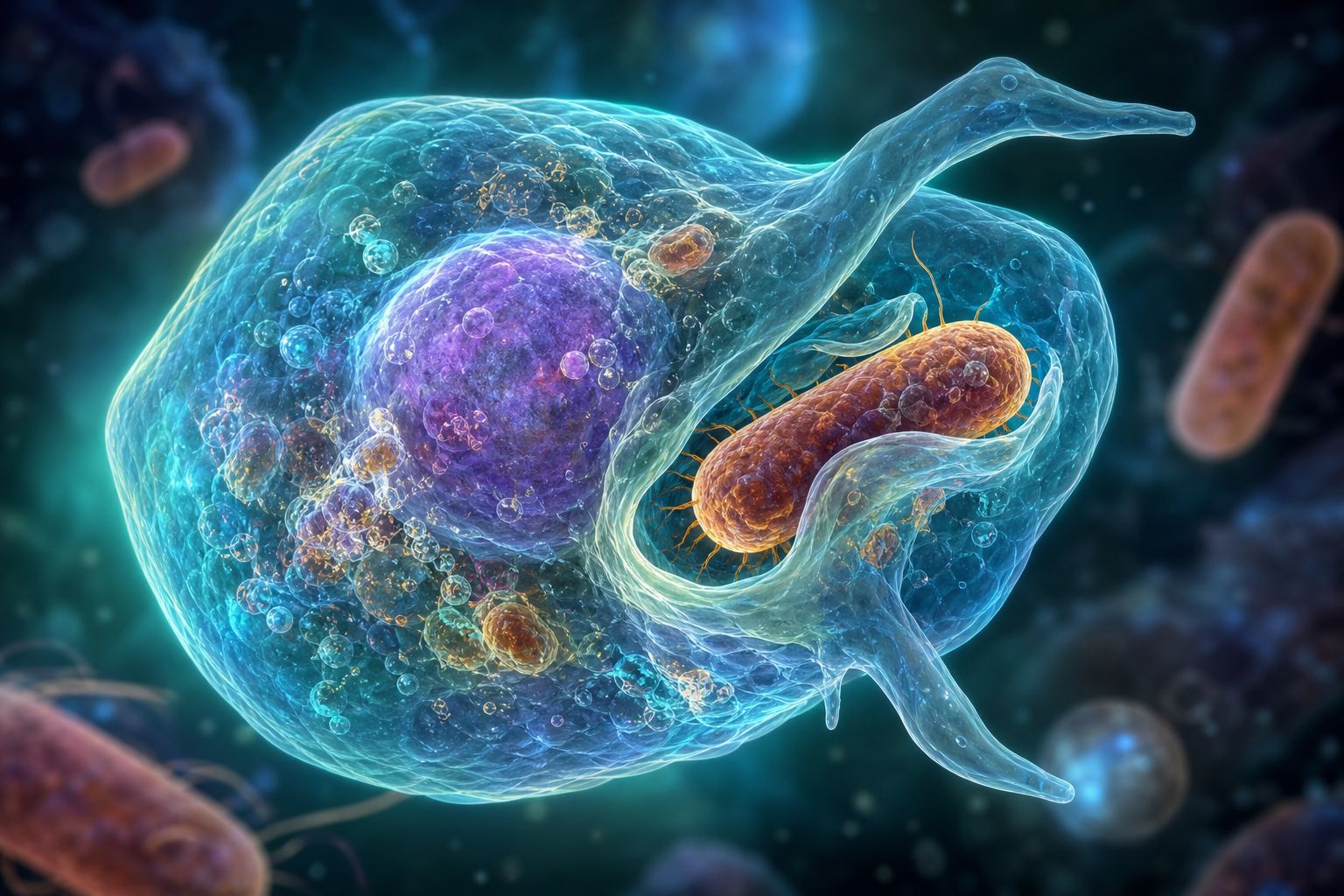
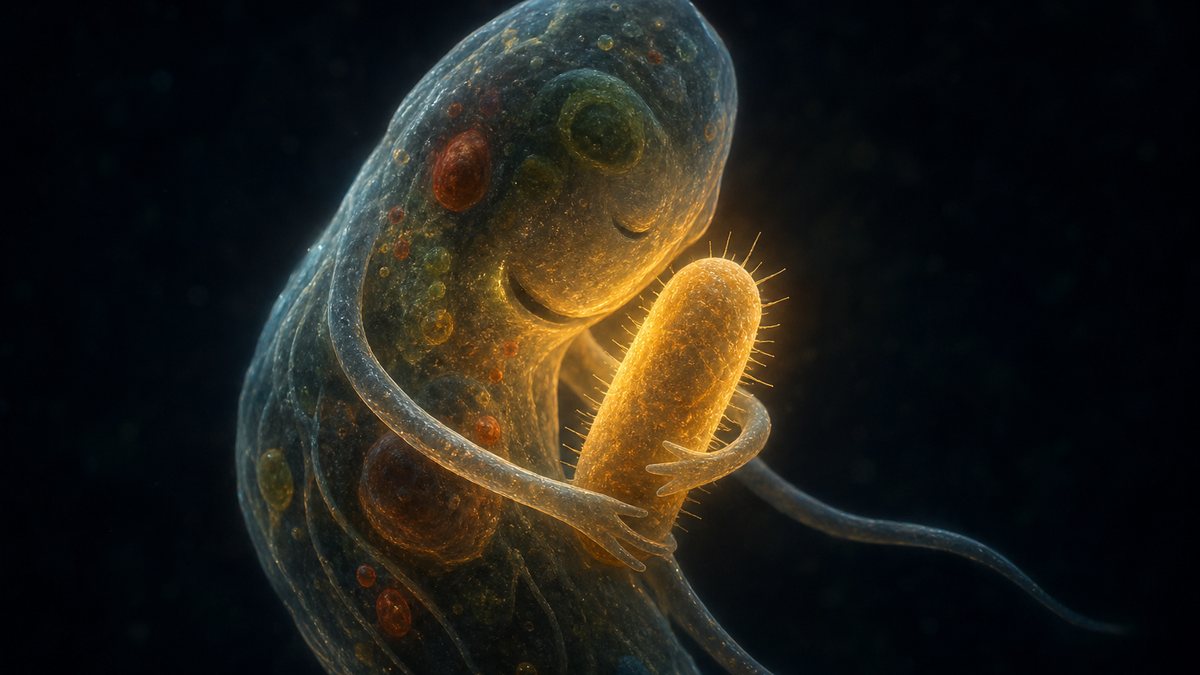
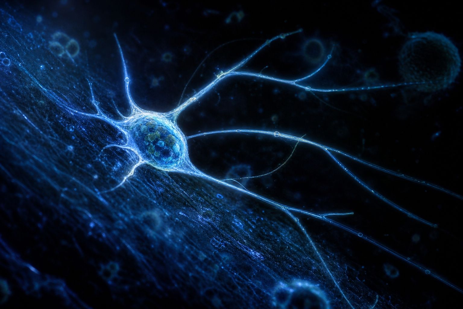
Arms on glass An artist’s impression of a microbe reaching out thin arms. AI-generated still.
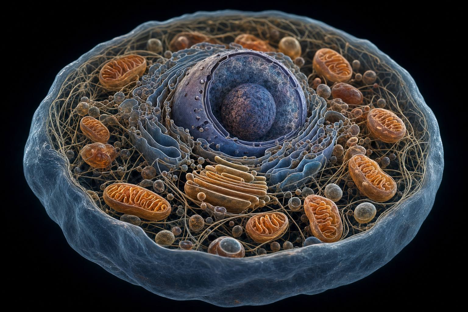
Inside a complex cell A complex cell holds a thousand times the volume of a bacterium, or more, and keeps its parts in separate compartments. AI-generated still.
  • Two kinds of cell
  • Bacteria living inside cells
  • The search for the host
  • Arms that crawl
Coming up
Prologue

A microbe that crawls

University of Vienna (30 September 2026): Philipp Radler and colleagues in Christa Schleper’s group, with JAMSTEC (Japan), IST Austria and HZI Braunschweig, report in Nature that two Asgard archaea, a Lokiarchaeon cultivated in Vienna and a Heimdallarchaeon cultivated in Japan, attach to surfaces and crawl using dynamic protrusions. Their cells are about 1,000 times smaller in volume than a human cell. The release calls Asgard archaea the closest known microbial relatives of eukaryotes.

This new study is the hook; chapter IV returns to it. The rest of the film is long-established biology.

The 3D scenes are simplified teaching models, not to scale.

Sources: University of Vienna (30 Sep 2026): Unexpected dynamics of ancient microbes discovered (Radler et al., Nature, doi 10.1038/s41586-026-11063-9)
Chapter I · Two kinds of cell

Every living thing is made of cells, and cells come in two basic designs.

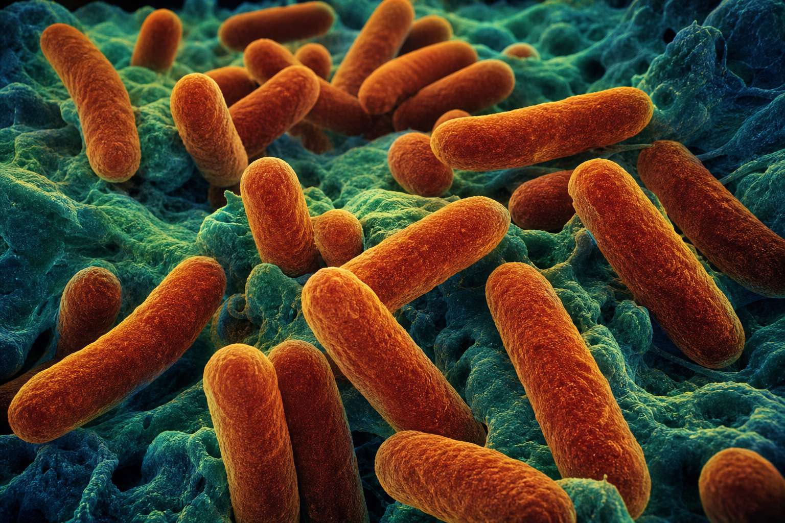
Bacteria and archaea are simple cells. Many are about a micrometre thick, and their DNA lies loose inside, with no nucleus around it.

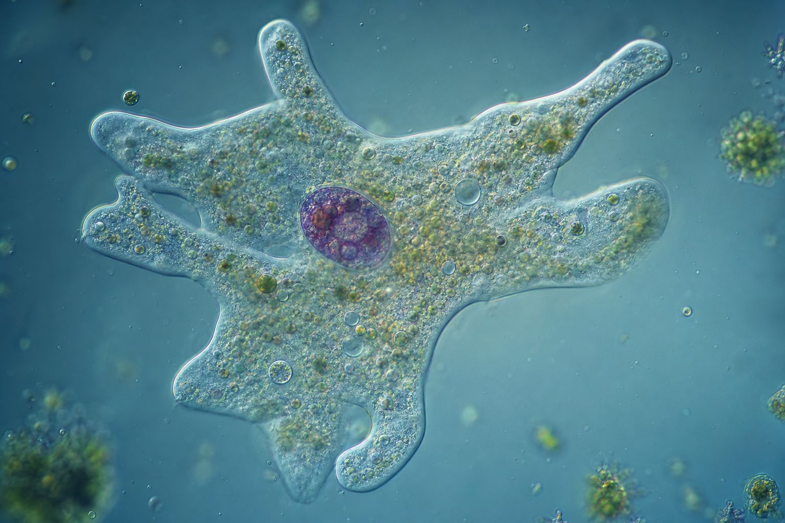
Plants, animals, fungi and single-celled organisms such as amoebas are built from complex cells, called eukaryotes. They keep their DNA inside a nucleus, wrapped in its own membrane.

Eukaryotic cells are usually much larger, and full of smaller compartments called organelles. Switch between the two designs.

Simple cells Rod-shaped bacteria, in the style of an electron micrograph. AI-generated still.
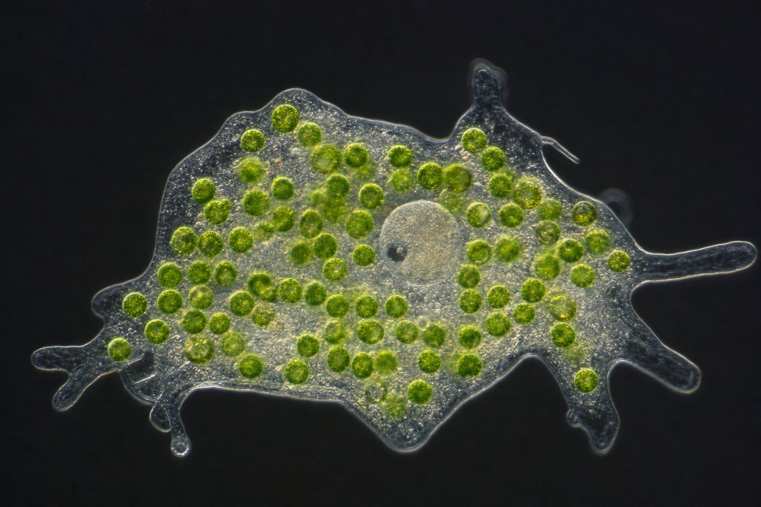
A complex cell An amoeba: one cell, with a nucleus. AI-generated still.
  • Prokaryotes (bacteria, archaea): no nucleus, no mitochondria
  • Eukaryotes: nucleus and organelles
The two designs
Chapter I · Two kinds of cell

Prokaryotes and eukaryotes

VAAM: prokaryotes (bacteria and archaea) have no nucleus, so their DNA is not enclosed in an envelope, and they lack mitochondria and chloroplasts; many are about one micrometre thick. Biology 1010 (13.2): life is divided into prokaryotes and eukaryotes, and eukaryotes include plants, animals, fungi and protists.

Try it: show the simple cell, the complex cell, or both.

The 3D scenes are simplified teaching models, not to scale.

Sources: VAAM, Association for General and Applied Microbiology: What are microbes?; Biology 1010 (OpenStax-based, Utah Education Network) 13.2: Eukaryotic Origins

Two kinds of organelle matter most to this story. Mitochondria use oxygen to release energy from food.

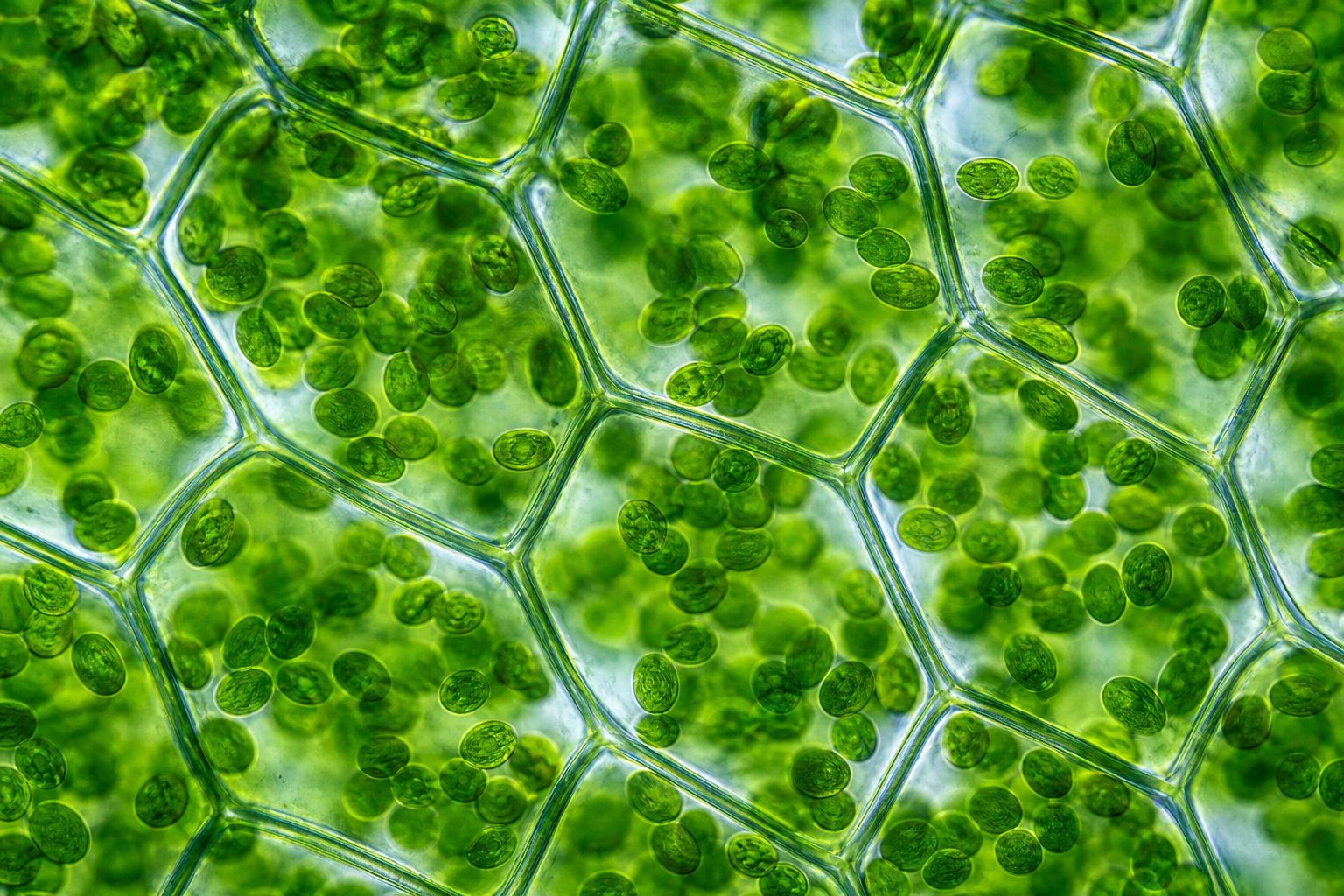
Plant and algal cells also have chloroplasts, which capture sunlight in photosynthesis. Compare an animal cell with a plant cell.

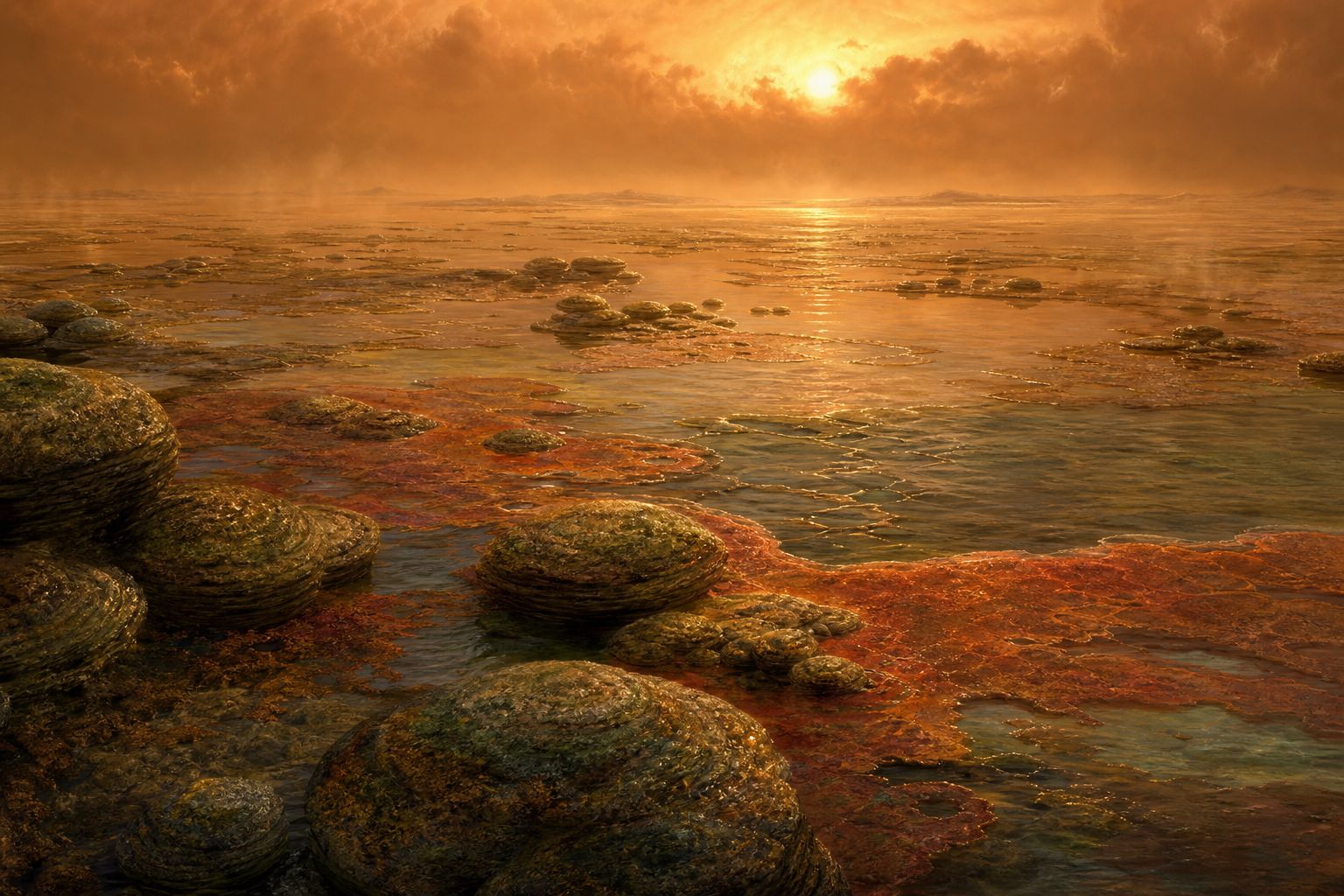
Simple cells appear about three and a half billion years ago. For well over a billion years after that, every cell on Earth was simple.

Complex cells appear about two billion years ago. Where did their nucleus and organelles come from?

1–10 µm
long: about the size of a bacterium
Mitochondria
Green compartments Leaf cells crowded with chloroplasts. AI-generated still.
  1. ~3.5 billion years ago simple cells
  2. ~2 billion years ago complex cells
  3. today both designs still thrive
Life on Earth
A world of microbes An ancient shallow sea with microbial mats; no animals or plants yet. AI-generated still.
Chapter I · Two kinds of cell

Organelles and deep time

Biology 1010 (13.2): prokaryotes appear about 3.5 billion years ago and eukaryotes about 2.1 billion years ago; mitochondria are 1 to 10 micrometres long. University of Vienna (2026): current models place the first eukaryotes around 2 billion years ago.

Dates this old come with wide error margins; “about” matters.

The 3D scenes are simplified teaching models, not to scale.

Sources: Biology 1010 (OpenStax-based, Utah Education Network) 13.2: Eukaryotic Origins; University of Vienna (30 Sep 2026): Unexpected dynamics of ancient microbes discovered (Radler et al., Nature, doi 10.1038/s41586-026-11063-9)
Chapter II · Cells within cells

Look closely at a mitochondrion and it looks surprisingly like a bacterium.

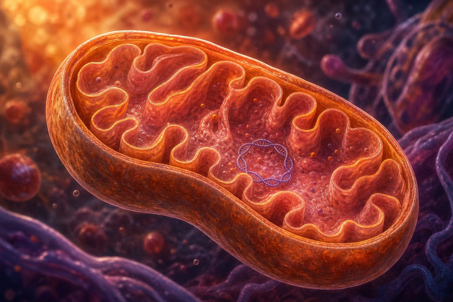
It has two membranes. Inside, it carries its own small loop of DNA, circular like a bacterium’s, separate from the DNA in the nucleus.

It makes some of its own proteins on its own ribosomes, the cell’s protein-making machines, which resemble bacterial ones, and it starts them with the same chemical signal that bacteria use.

New mitochondria come only from old ones, which divide in two much as a bacterium does.

Even an antibiotic agrees. Streptomycin, which stops bacteria making proteins, also stops mitochondria, but not the rest of the cell.

Cutaway Folded inner membrane and a ring of DNA. AI-generated still.
16,569
base pairs in one small ring
Human mitochondrial DNA
  • Two membranes
  • Circular DNA, like bacteria
  • Bacteria-sized ribosomes (70S)
  • Same protein start signal as bacteria (fMet)
Bacteria-like clues
  • Divide like bacteria (binary fission)
  • Never made from scratch by the cell
More clues
  • Streptomycin: stops bacteria and mitochondria
  • Diphtheria toxin: stops the cell’s own ribosomes, not theirs
The antibiotic test
Chapter II · Cells within cells

Why mitochondria look like bacteria

Kimball 18.11: mitochondria have their own single circular DNA molecule without histones (16,569 base pairs in humans), their own bacteria-like protein-making machinery, start proteins with fMet as bacteria do, arise only from pre-existing mitochondria, and are blocked by streptomycin, which spares the eukaryotic cytoplasm. BioNinja: double membrane, 70S ribosomes and division resembling binary fission.

Try it: watch it divide.

The 3D scenes are simplified teaching models, not to scale.

Sources: LibreTexts, Biology (Kimball) 18.11: Endosymbiosis; BioNinja: Endosymbiosis (evidence)

In 1967 the biologist Lynn Margulis, then publishing as Lynn Sagan, set out a bold explanation.

Mitochondria, she argued, began as free-living bacteria that came to live inside another cell. Over many generations the guest became part of its host.

The idea is called endosymbiosis, which means living together, one inside the other.

More than a dozen journals turned her paper down before the Journal of Theoretical Biology accepted it. Today the idea that mitochondria and chloroplasts began as bacteria is widely accepted.

On the Origin of Mitosing CellsLynn Sagan (Margulis), Journal of Theoretical Biology
The 1967 paper
A guest moves in A large cell wrapping around a smaller bacterium. AI-generated still.
  • endo: inside
  • sym: together
  • biosis: living
The word
12+
journals rejected it, partly because it crossed many fields
Hard to publish
It still happens Some single-celled hunters today keep live algae inside them and live partly on what the algae make. AI-generated still.
Chapter II · Cells within cells

Margulis and endosymbiosis

Understanding Science (UC Berkeley): in 1967 Lynn Margulis, then Lynn Sagan, published “On the Origin of Mitosing Cells”; more than a dozen journals rejected it, not because the science was poor but because it did not fit one field, before the Journal of Theoretical Biology accepted it; a book followed in 1970. Biology 1010 (13.2): the endosymbiotic hypothesis is now widely accepted.

Try it: replay the merger.

The 3D scenes are simplified teaching models, not to scale.

Sources: Understanding Science, UC Berkeley: Cells within cells, the community reacts; Biology 1010 (OpenStax-based, Utah Education Network) 13.2: Eukaryotic Origins

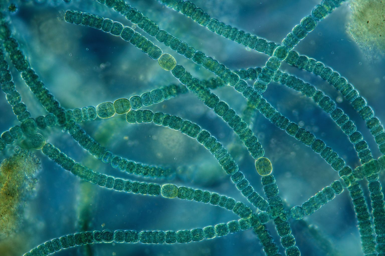
The same story explains chloroplasts. They descend from cyanobacteria, the blue-green bacteria that photosynthesise.

Like mitochondria, chloroplasts carry their own circular DNA and make proteins on bacteria-like ribosomes.

The chloroplast came second. Every known complex cell has mitochondria, or stripped-down organelles descended from them, but only some have chloroplasts.

So a cell that already had mitochondria took in a cyanobacterium. That is why a leaf cell carries two kinds of former bacteria, and yours carry one.

Cyanobacteria Chains of small blue-green cells. AI-generated still.
121,024
base pairs, about seven times a human mitochondrion’s
A liverwort’s chloroplast DNA
  1. first a bacterium becomes the mitochondrion
  2. later a cyanobacterium becomes the chloroplast
Order of arrival
Two former bacteria Each green oval descends from a once free-living cyanobacterium. AI-generated still.
Chapter II · Cells within cells

Chloroplasts

Kimball 18.11: chloroplasts descend from an endosymbiotic cyanobacterium that lived inside a eukaryotic cell that already had mitochondria; they have their own circular DNA (121,024 base pairs in the liverwort Marchantia) and bacteria-like protein synthesis. Biology 1010 (13.2): mitochondria likely came before plastids, because all eukaryotes have mitochondria or mitochondria-like organelles but only some have plastids.

Try it: replay the merger.

The 3D scenes are simplified teaching models, not to scale.

Sources: LibreTexts, Biology (Kimball) 18.11: Endosymbiosis; Biology 1010 (OpenStax-based, Utah Education Network) 13.2: Eukaryotic Origins

Chapter III · Who was the host?

That leaves a question. If the mitochondrion was the guest, who was the host?

Many genes that copy and read the DNA in our nucleus are closely related to those of archaea, the other kind of simple cell. So the host was probably an archaeon.

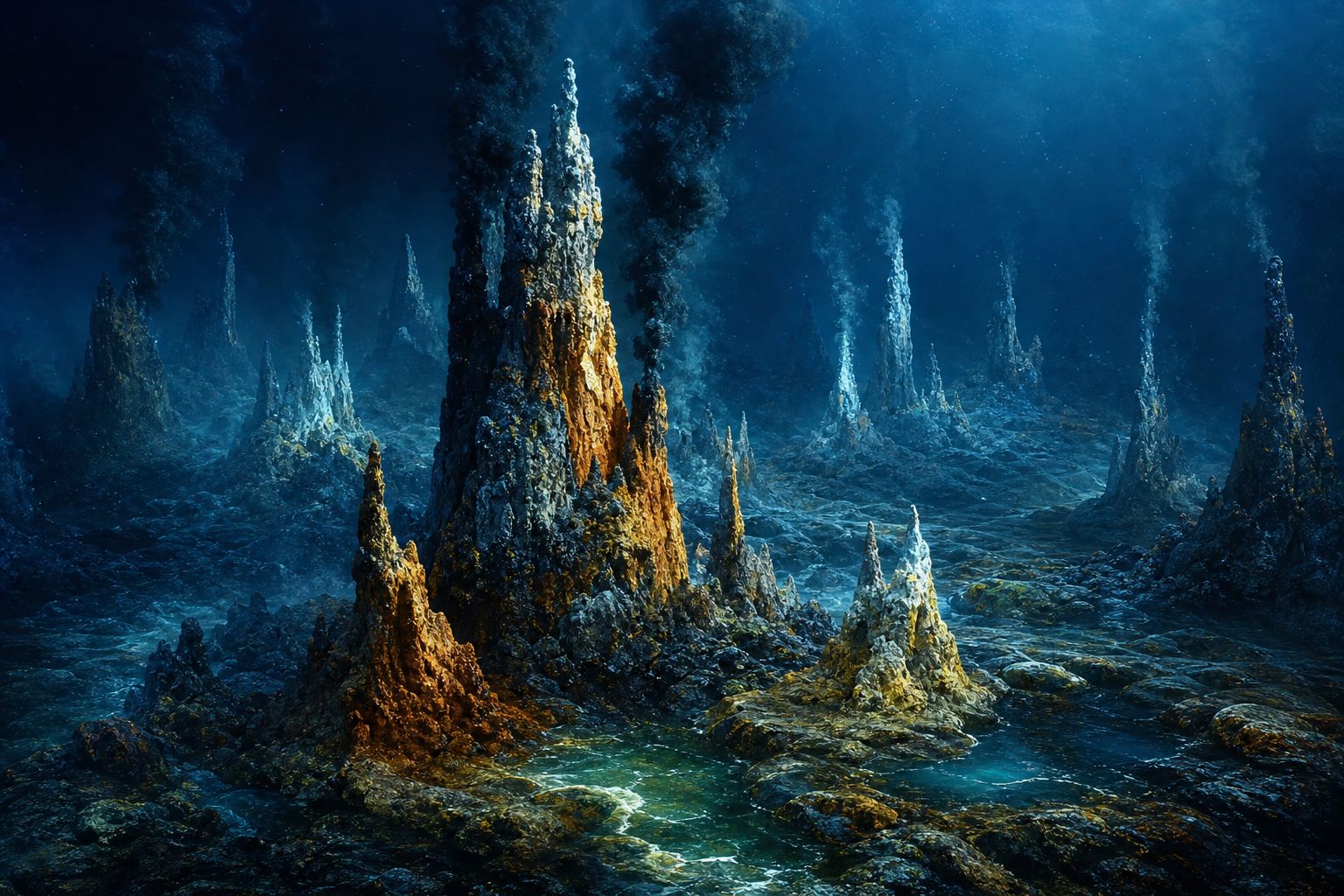
In 2015 a team led by Thijs Ettema at Uppsala University in Sweden found DNA from a new group of archaea in sea-floor mud near a hydrothermal vent field called Loki’s Castle, in the Arctic seas between Greenland and Norway.

They called it Lokiarchaeota. The wider group, named after the home of the Norse gods, is the Asgard archaea.

Their genes put them closer to complex cells than any other known microbe, and many researchers think eukaryotes branched off from within them.

  • Guest: a bacterium (the mitochondrion)
  • Host: ?
The puzzle
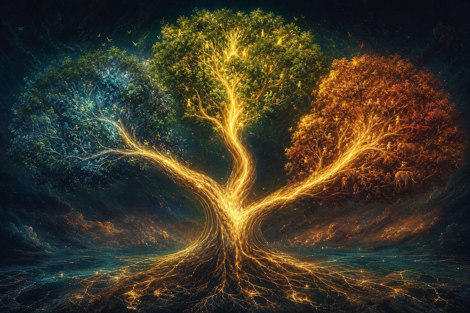
Three great branches Bacteria, archaea and eukaryotes. AI-generated still.
Vent field Mineral chimneys on a dark sea floor, like those near Loki’s Castle. AI-generated still.
2015
genes first, no living cells yet
Found from DNA alone
  • Three domains: eukaryotes a separate branch
  • Two domains: eukaryotes grew from within the archaea
Domains: the biggest branches of life
Chapter III · Who was the host?

Finding the host

Biology 1010 (13.2): many nuclear genes of eukaryotes and their replication and expression machinery appear closely related to archaea, while energy-harvesting genes came from bacteria. Kimball 18.11 describes the host as an archaeal cell. Wikipedia (Promethearchaeum): Lokiarchaeota were reported in 2015 by Thijs Ettema’s team at Uppsala University from near the Loki’s Castle vent. Earth.com (2025): Asgard archaea, named from Norse mythology, were identified in 2015 from gene fragments; some scientists argue eukaryotes evolved from within them, which would leave two primary domains.

The tree on screen is simplified; branch lengths mean nothing.

The 3D scenes are simplified teaching models, not to scale.

Sources: Biology 1010 (OpenStax-based, Utah Education Network) 13.2: Eukaryotic Origins; LibreTexts, Biology (Kimball) 18.11: Endosymbiosis; Wikipedia: Promethearchaeum (Lokiarchaeota 2015; Imachi et al., Nature, January 2020); Earth.com (24 Mar 2025): Tiny microbes may have given rise to complex life

But genes in the mud are not a living cell. Nobody had seen what an Asgard archaeon looked like.

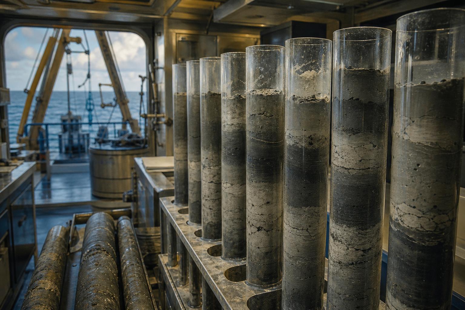
A Japanese team led by Hiroyuki Imachi took about twelve years to grow one, from sea-floor mud collected about two and a half kilometres deep off Shikoku.

Reported in 2020, it would only grow alongside partner microbes. Its cells, about half a micrometre wide, sprouted long, branching arms.

Its DNA held about eighty genes once thought to belong only to complex cells. A real Asgard archaeon was finally under the microscope.

Sea-floor mud Sediment cores like those that held the microbe. AI-generated still.
~12 years
to grow it, publish in Nature, January 2020
Patience
~0.55 µm
cell width; arms about 5 µm long
Promethearchaeum
80
“eukaryotic signature” genes
Borrowed-looking genes
Chapter III · Who was the host?

The first one grown

Wikipedia (Promethearchaeum): Hiroyuki Imachi and colleagues reported in Nature in January 2020 the culture of Promethearchaeum syntrophicum from deep-sea sediment of the Nankai Trench off Shikoku, at about 2,500 m, after about 12 years; it grows only with partners (a Methanogenium archaeon and a Halodesulfovibrio bacterium), its cells are about 550 nm across, its tentacle-like protrusions, some branched, average about 5 µm long, and its genome holds 80 eukaryotic signature genes. University of Vienna (2022): the first images of Asgard cells came from this Japanese group in 2020.

The 3D scenes are simplified teaching models, not to scale.

Sources: Wikipedia: Promethearchaeum (Lokiarchaeota 2015; Imachi et al., Nature, January 2020); University of Vienna via idw (21 Dec 2022): On the trail of the origin of complex life (Rodrigues-Oliveira et al., Nature, doi 10.1038/s41586-022-05550-y)

Put the pieces together and you get the leading model.

About two billion years ago, a cell related to today’s Asgard archaea joined with a bacterium. The bacterium lived on inside it and became the mitochondrion.

Over a very long time the partners came to depend on each other. Many proteins a mitochondrion needs are now made from genes in the nucleus.

How the bacterium got inside, and what each side gained, is still debated. This is the model the evidence favours, not a film of what happened.

  • Host: an Asgard-like archaeon
  • Guest: a bacterium
  • Later, in some lines: a cyanobacterium
The partners
Two billion years of layers Two billion years is most of the history of life; the first animals turn up only in the last fifth of it. AI-generated still.
2 genomes
a mitochondrion relies on its own DNA and the nucleus
Shared control
  • Swallowed, or slowly wrapped by arms?
  • Which came first: nucleus or mitochondrion?
  • What each partner traded
Still debated
Chapter III · Who was the host?

Two cells become one

University of Vienna (2026): current models suggest the first eukaryotes arose around 2 billion years ago from a fusion of a bacterium with an Asgard-like ancestor. Kimball 18.11: nuclear genes encode many of the proteins in mitochondria and chloroplasts, so the organelles can arise only from pre-existing ones. Earth.com (2025): researchers propose that an Asgard archaeon may have used its tentacle-like arms to capture the bacterium that became the mitochondrion, a hypothesis.

Try it: replay the merger.

The 3D scenes are simplified teaching models, not to scale.

Sources: University of Vienna (30 Sep 2026): Unexpected dynamics of ancient microbes discovered (Radler et al., Nature, doi 10.1038/s41586-026-11063-9); LibreTexts, Biology (Kimball) 18.11: Endosymbiosis; Earth.com (24 Mar 2025): Tiny microbes may have given rise to complex life

Chapter IV · Arms that crawl

In 2022, Christa Schleper’s group in Vienna found that one Asgard archaeon is threaded with a network of actin filaments, a scaffold like those seen before only in complex cells.

In complex cells, actin can push out the edge of a cell so that it crawls. Some human immune cells move this way.

Now Philipp Radler and colleagues have filmed living cells of two Asgard strains, kept free of oxygen: one from the Loki branch, grown in Vienna, and one from the Heimdall branch, grown in Japan.

The cells changed shape about every minute, stretched arms up to twenty times their body length, stuck to the glass and crawled.

When the team added drugs that block actin, the cells lost control of their arms and stopped moving. Try it.

actin
filament network inside Lokiarchaeum ossiferum
Nature, 2022
Crawling, the complex way Complex cells such as amoebas push out their edges to move. AI-generated still.
  • Lokiarchaeon: grown in Vienna
  • Heimdallarchaeon: grown in Japan (JAMSTEC)
The two strains
20×
arm length versus body
~1 min
between big shape changes
Nature, 2026
  • Normal: arms reach, attach, pull
  • Actin blocked: arms out of control, no crawling
The test
Chapter IV · Arms that crawl

Arms that crawl

University of Vienna via idw (2022): Schleper’s group, with ETH Zürich, grew Lokiarchaeum ossiferum from sediment off Piran, Slovenia, and found an extensive actin network previously seen only in eukaryotes. University of Vienna (2026): Radler et al. used live-cell microscopy in oxygen-free conditions on a Lokiarchaeon and a Heimdallarchaeon; cells changed shape drastically about every minute, protrusions reached up to 20 times the cell body, cells attached and crawled, a motility previously seen only in complex cells such as human immune cells; actin inhibitors stopped the movement.

Try it: add the actin inhibitor.

The 3D scenes are simplified teaching models, not to scale.

Sources: University of Vienna via idw (21 Dec 2022): On the trail of the origin of complex life (Rodrigues-Oliveira et al., Nature, doi 10.1038/s41586-022-05550-y); University of Vienna (30 Sep 2026): Unexpected dynamics of ancient microbes discovered (Radler et al., Nature, doi 10.1038/s41586-026-11063-9)
What does this tell us? The authors suggest that crawling, a hallmark of complex cells, may be older than complex cells themselves.

If so, the archaeal host may already have had a working actin skeleton before it took in the bacterium that became the mitochondrion.

But today’s Asgard archaea are cousins, not ancestors. They have kept evolving for two billion years too, so they are a living model, not a time machine.

And only a handful have ever been grown and watched alive, so the conclusions rest on very few kinds of cell.

  • Crawling may predate complex cells
  • Actin may have mattered before the merger
What the study suggests
Arms before partners? An idea to test, not a finding. AI-generated still.
  • Today’s archaea are not the ancestor
  • Two strains, not the whole group
  • How the merger actually happened
What it cannot show
2020, 2023
first two Asgard specimens cultivated, per the University of Vienna
Grown in labs
Chapter IV · Arms that crawl

What it means, and what it doesn’t

University of Vienna (2026): the authors suggest that complex cell motility may be much older than assumed, and that these traits may have mattered even before the first eukaryotes and the symbiosis that gave rise to mitochondria; the release frames this as a possibility (“may”, “suggests”). The first two Asgard specimens were cultivated in 2020 and 2023, at JAMSTEC and in Schleper’s lab. Martin Pilhofer (ETH Zürich), quoted by Earth.com (2025): “We still have a lot of unanswered questions about Asgard archaea, especially regarding their relation to eukaryotes.”

“Cousins, not ancestors” is the standard caution when living species are used to picture the past.

The 3D scenes are simplified teaching models, not to scale.

Sources: University of Vienna (30 Sep 2026): Unexpected dynamics of ancient microbes discovered (Radler et al., Nature, doi 10.1038/s41586-026-11063-9); Earth.com (24 Mar 2025): Tiny microbes may have given rise to complex life

Epilogue

Every breath you take carries oxygen to the mitochondria in your cells, descendants of a bacterium that moved in about two billion years ago.

Complex life seems to have begun not with one cell defeating another, but with two very different cells living together.

How a small archaeon first took in its partner is still one of biology’s great open questions.

  1. ~2 billion years ago archaeon and bacterium merge
  2. 1967 Margulis’s endosymbiosis paper
  3. 2015 Lokiarchaeota found from DNA
  4. 2020 first Asgard archaeon grown
  5. 2026 Asgard archaea filmed crawling
The cell inside the cell: a timeline
Living together Two very different cells, one future. AI-generated still.
  • How did the bacterium get inside?
  • Where did the nucleus come from?
Open questions