In October 2026 researchers at Washington University in St. Louis reported that picobirnaviruses, common gut viruses long thought to infect animal cells, infect a gut bacterium instead. This film explains the established biology behind that news: what a virus is, the discovery of bacteriophages by Twort and d'Hérelle, the anatomy of phage T4, the lytic cycle from attachment to the burst, why one phage makes a visible plaque, the lysogenic cycle of phage lambda, the Hershey–Chase experiment that showed genes are DNA, CRISPR as bacterial immunity and the genetic scissors it led to, how many phages there are in the sea and the gut, and the history and present limits of phage therapy.
Interactive 3D film10 min1892 to 2026Published 3 November 2026
How the viruses that hunt bacteria work, and what they taught us.
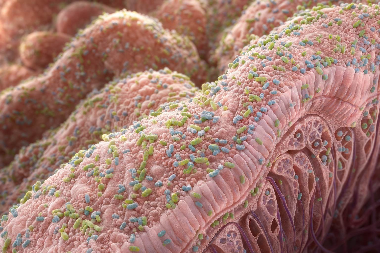
Your gut holds trillions of bacteria. And they have enemies of their own: viruses that can infect bacteria and nothing else.
In October 2026, researchers reported that one of the commonest viruses in the human gut, long thought to infect our own cells, seems to infect a gut bacterium instead.
It is one study, and it needs confirming. But the viruses it points to, the bacteriophages, are among the best understood things in biology.
Here is how they work.
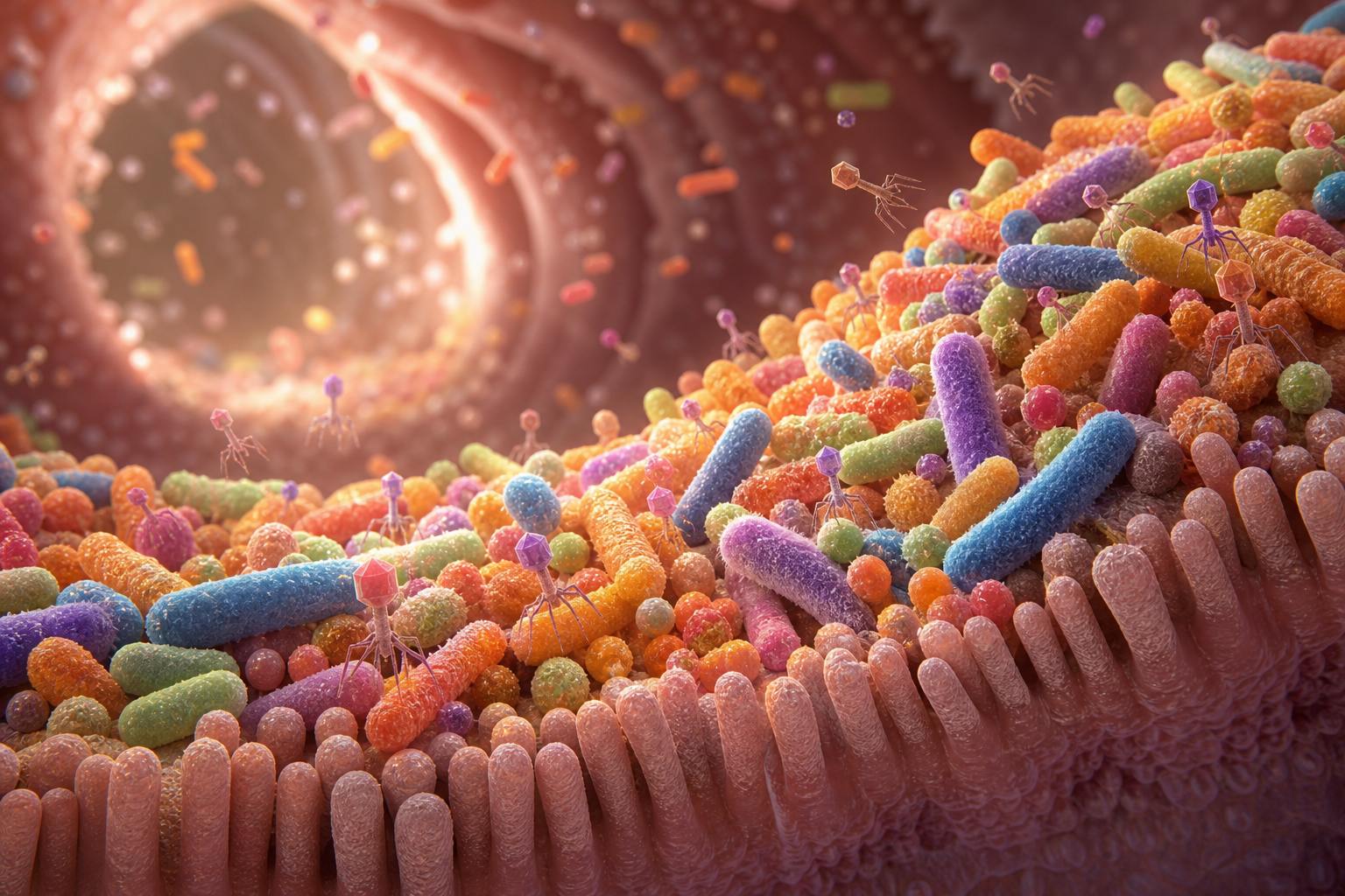
The gut Bacteria crowd the folds of the intestinal wall, a few thousandths of a millimetre across. AI-generated still.
Picobirnaviridae
found inside a newly named gut bacterium, Candidatus Anaerotignum faecalis
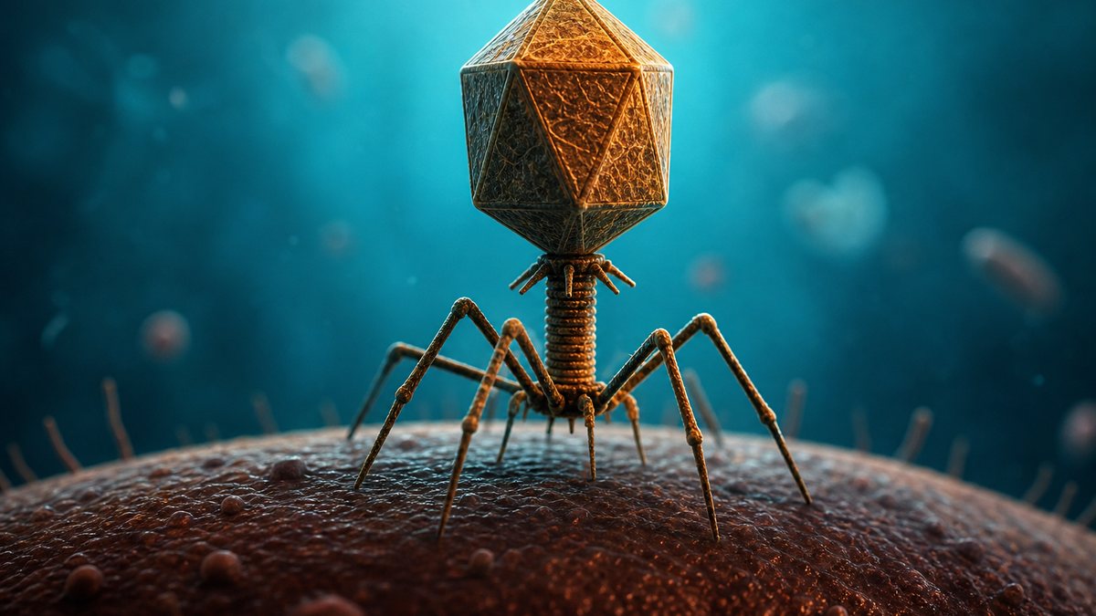
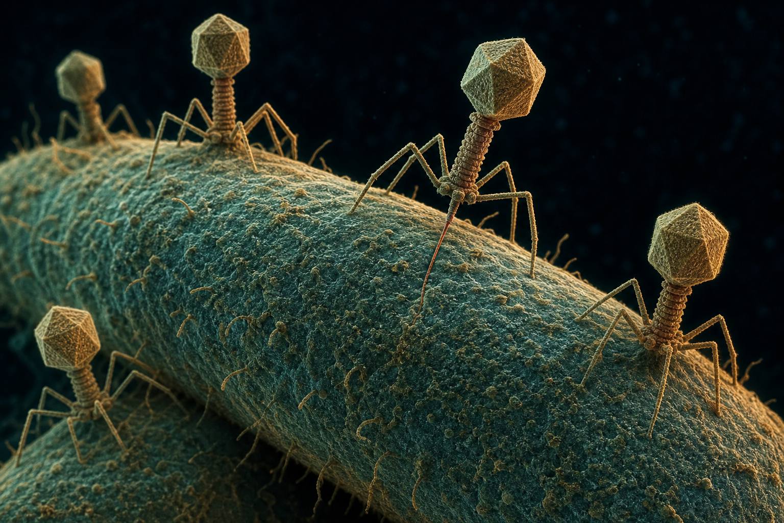
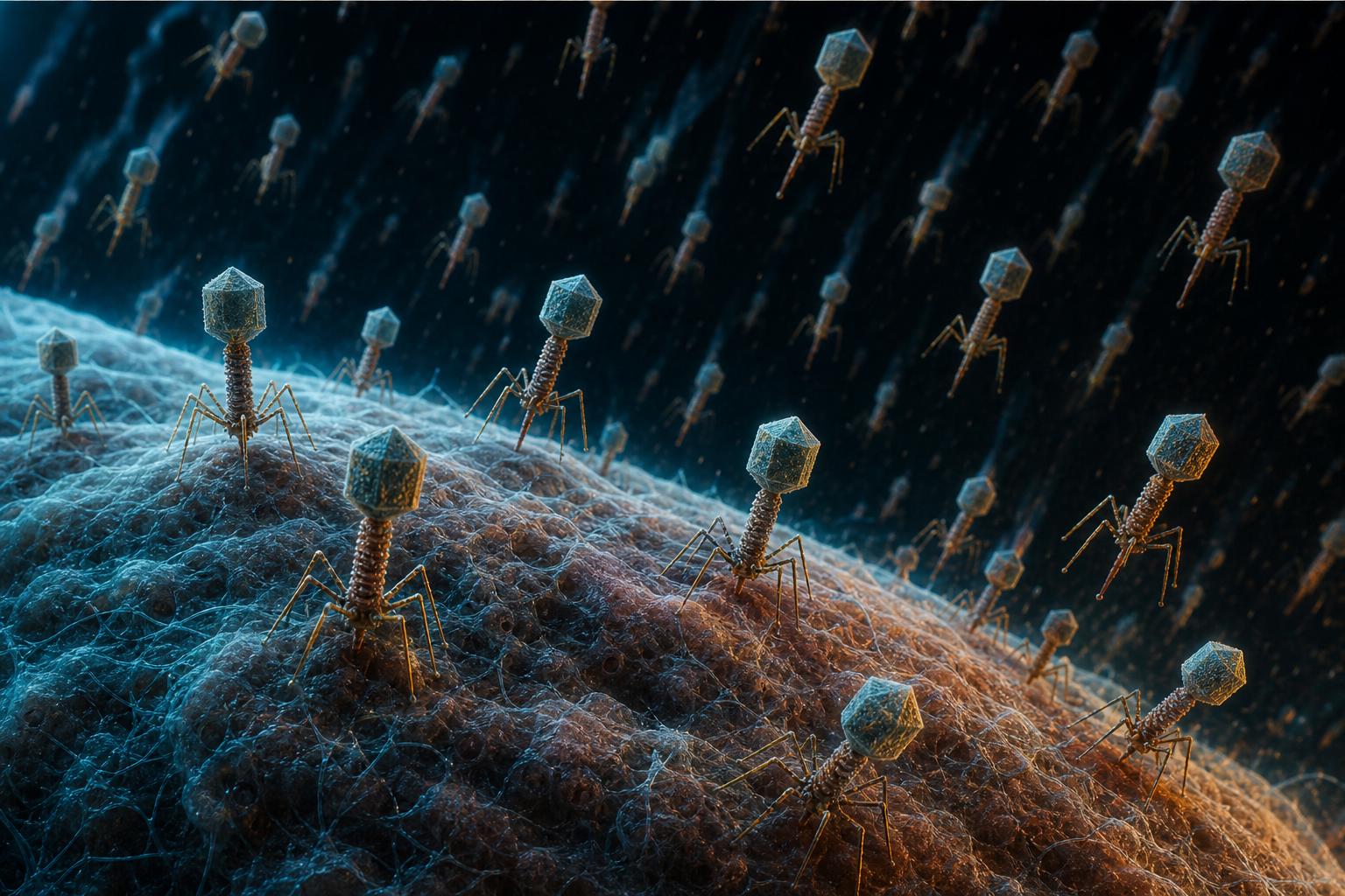
Washington University in St. Louis, in Nature Microbiology, 29 Oct 2026Bacteriophages Tailed phages land on a bacterium, in the style of an electron micrograph. AI-generated still.
Who discovered phages
How a phage kills, or hides
DNA, CRISPR and the oceans
Phages as medicine
Coming up
Prologue
A gut virus that hunts bacteria
WashU Medicine (29 October 2026): Tianyu Gan, David Wang and colleagues report in Nature Microbiology (“A human gut-associated picobirnavirus establishes the Picobirnaviridae as the third clade of RNA bacteriophages”) that picobirnaviruses, long thought to infect human gut cells, infect gut bacteria. In germ-free mice given human stool the virus multiplied; grown with gut bacteria without oxygen it multiplied for 20 days; antibiotics that killed the bacteria stopped it; imaging placed it inside a newly identified bacterium, Candidatus Anaerotignum faecalis. Wang cautions that finding high levels in patients with some diseases “does not prove the virus causes disease.”
This single new study is the hook. The rest of the film is long-established phage biology.
The 3D scenes are simplified teaching models, not to scale.
A virus is not a cell. It is a set of genes packed inside a shell of protein called a capsid. The genes are written either in DNA, or in RNA, a closely related molecule, but never in both.
On its own it does nothing at all. It can only copy itself inside a living cell, by taking over that cell's machinery.
Viruses infect every kind of life. The ones that infect bacteria are called bacteriophages, or phages for short.
Genes: DNA or RNA
A protein shell, the capsid
Some also wear a fatty envelope
What a virus is
~20–900 nm
most too small for a light microscope
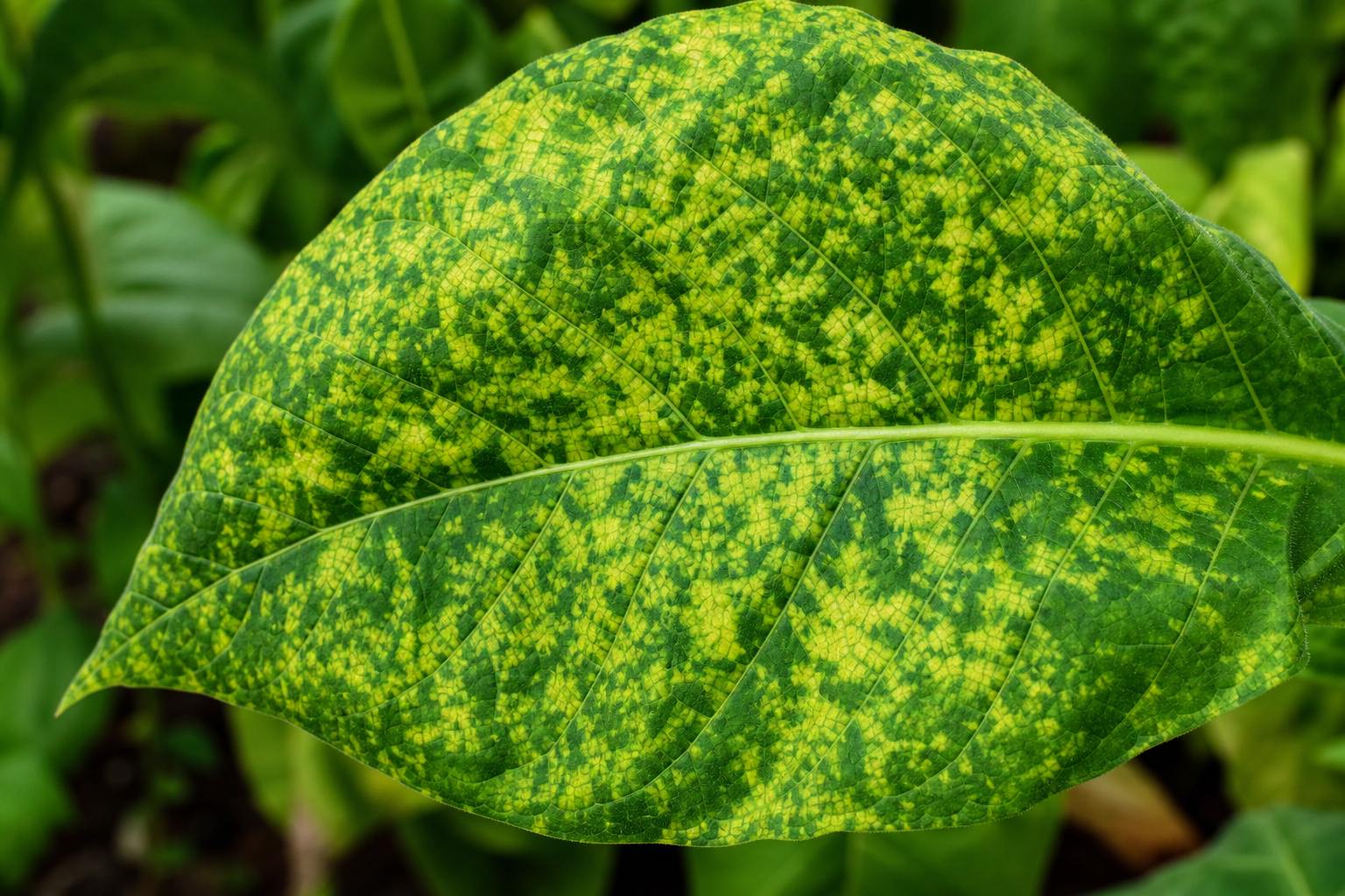
Size (OpenStax)Tobacco mosaic In 1892 the Russian botanist Dmitri Ivanovski found that this leaf disease passed through a filter too fine for bacteria; in 1899 Martinus Beijerinck called the agent a “virus”, Latin for poison. AI-generated still.A phage A head of protein full of DNA, on a tail with legs. AI-generated still.
Chapter I · The bacteria eaters
What is a virus?
OpenStax Microbiology 6.1: viruses are acellular, obligate intracellular parasites that cannot reproduce on their own; each has a single kind of nucleic acid, DNA or RNA, never both, inside a protein capsid, and some have a lipid envelope from the host; virions range from about 20 nm to about 900 nm. In 1892 Dmitri Ivanovski found that tobacco mosaic disease passed through a porcelain filter that removed bacteria; in 1899 Martinus Beijerinck concluded the agent was not a bacterium, which led to the term virus, Latin for poison. Complex phages have a polyhedral head and a sheath linking it to tail fibres.
The 3D scenes are simplified teaching models, not to scale.
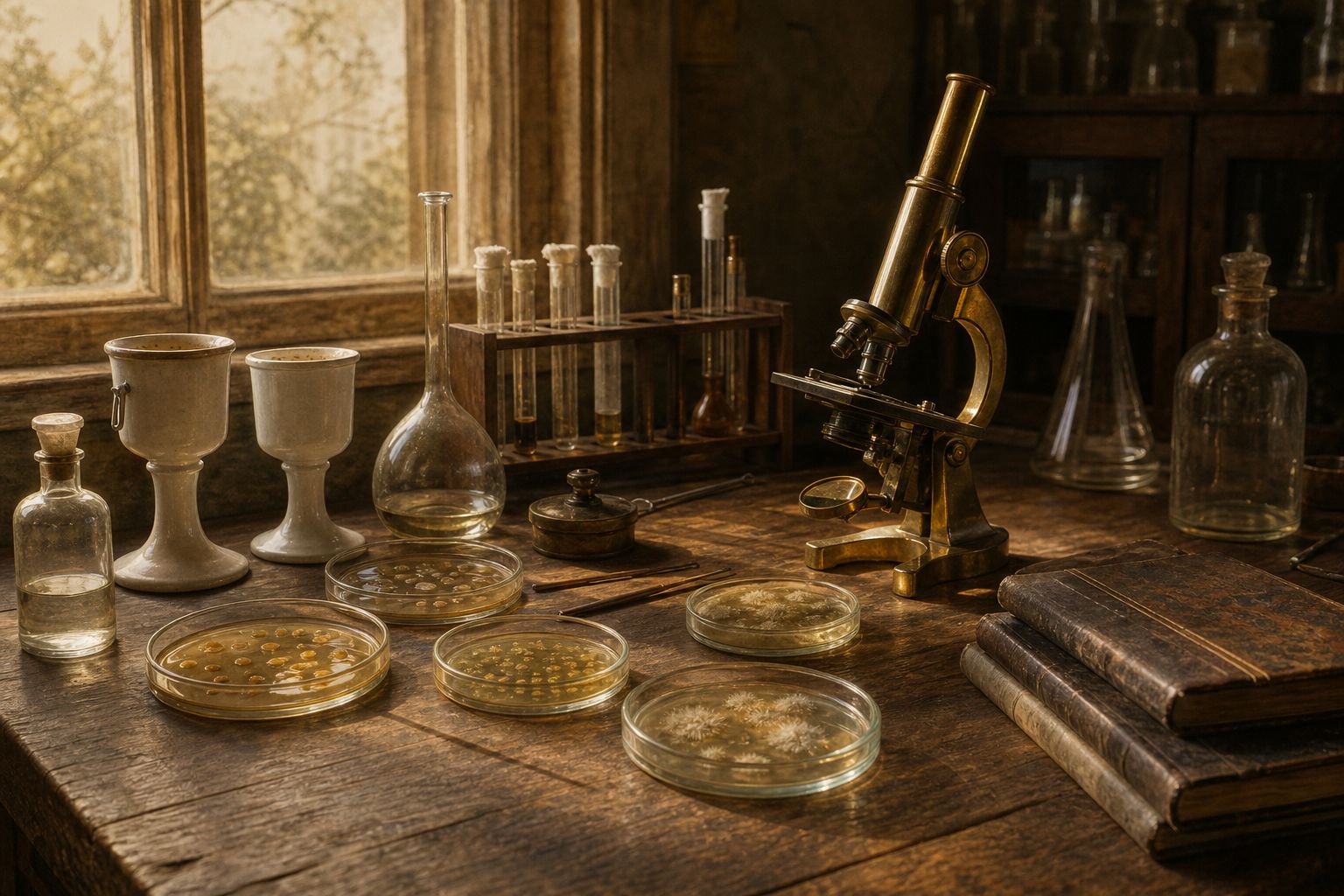
In 1915 the English bacteriologist Frederick Twort described, in The Lancet, glassy, transparent patches spreading through his bacterial cultures.
Whatever killed the bacteria passed through a porcelain filter, and could be passed from plate to plate.
In 1917 Félix d'Hérelle, at the Pasteur Institute in Paris, saw the same thing in bacteria from dysentery patients: clear spots where the bacteria had vanished.
He was sure it was an invisible microbe that ate bacteria, and named it the bacteriophage: the bacteria eater.
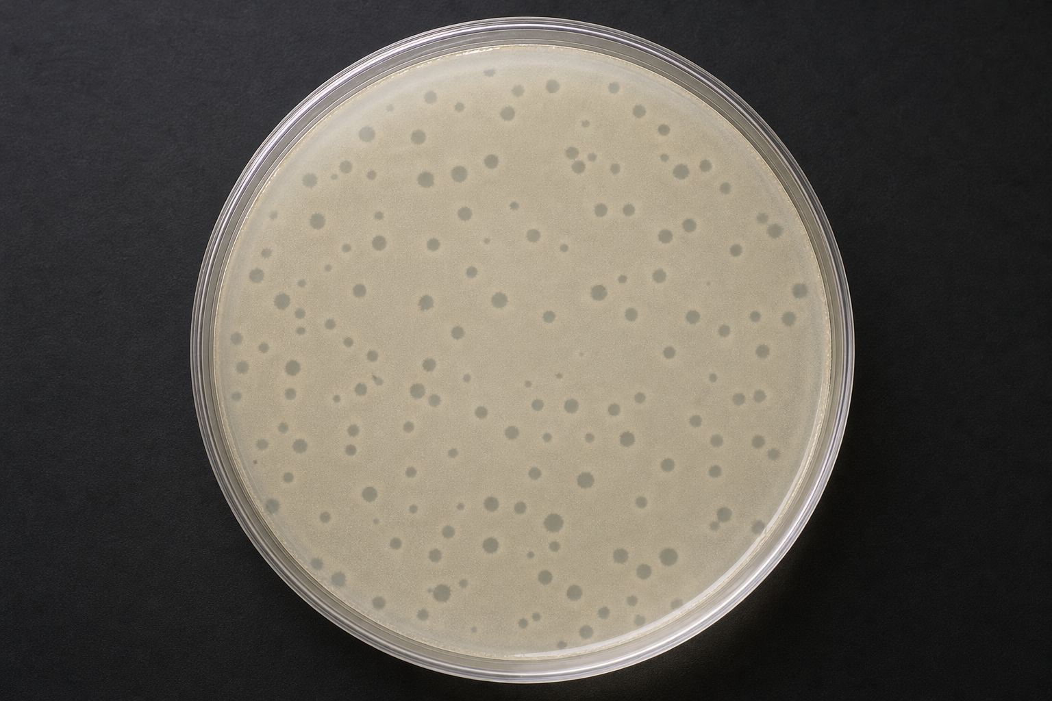
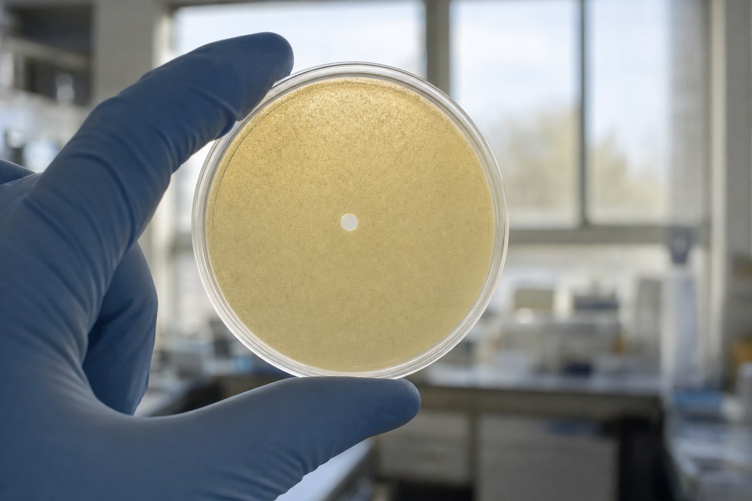
Those clear spots are called plaques. Each one starts from a single phage.
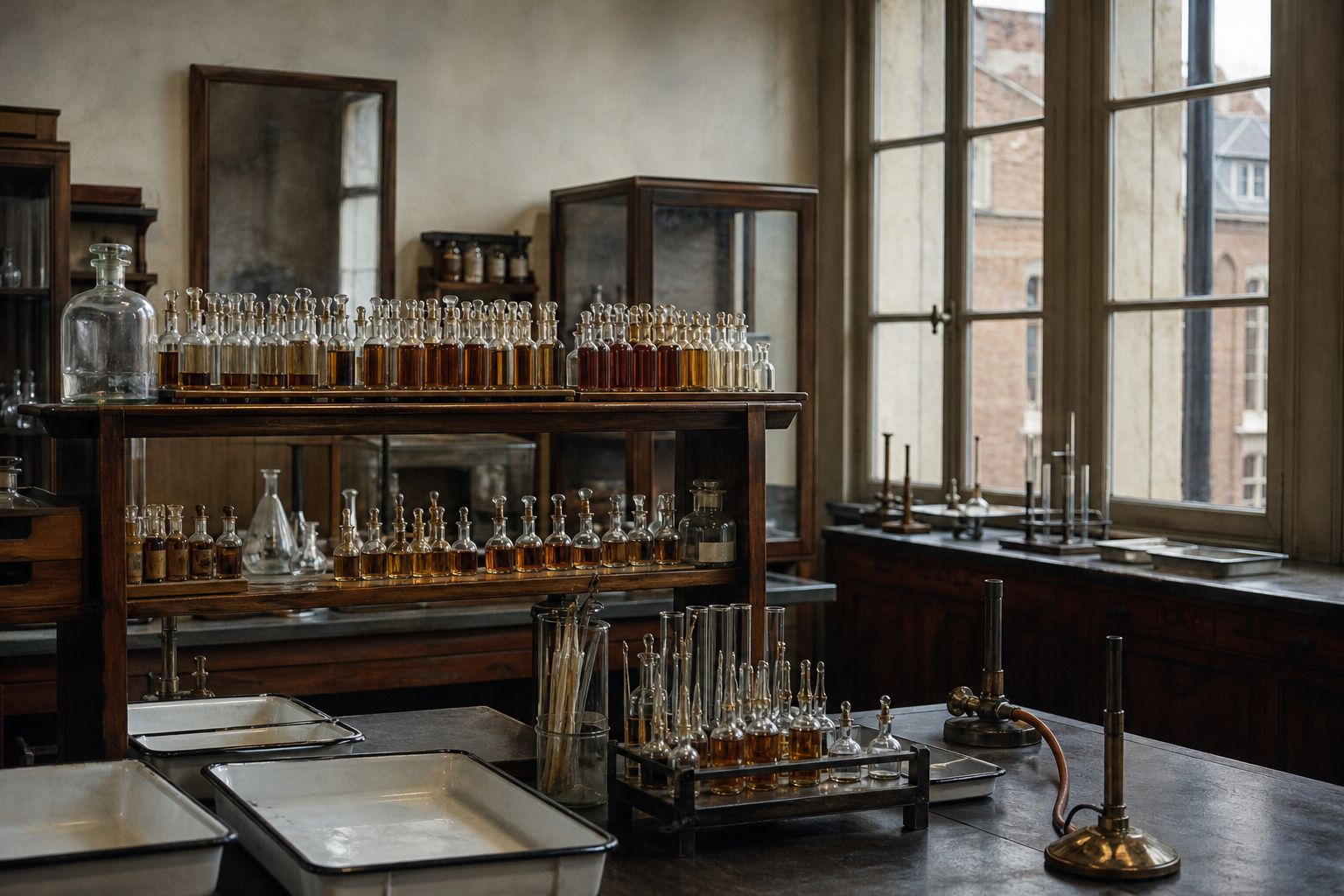
A wartime bench Agar plates, flasks and filter candles of the 1910s. Twort’s work stopped when he joined the Royal Army Medical Corps. AI-generated still.
1915 Twort, London: “glassy and transparent” patches
1917 d’Hérelle, Paris: “an invisible microbe antagonistic to dysentery bacilli”
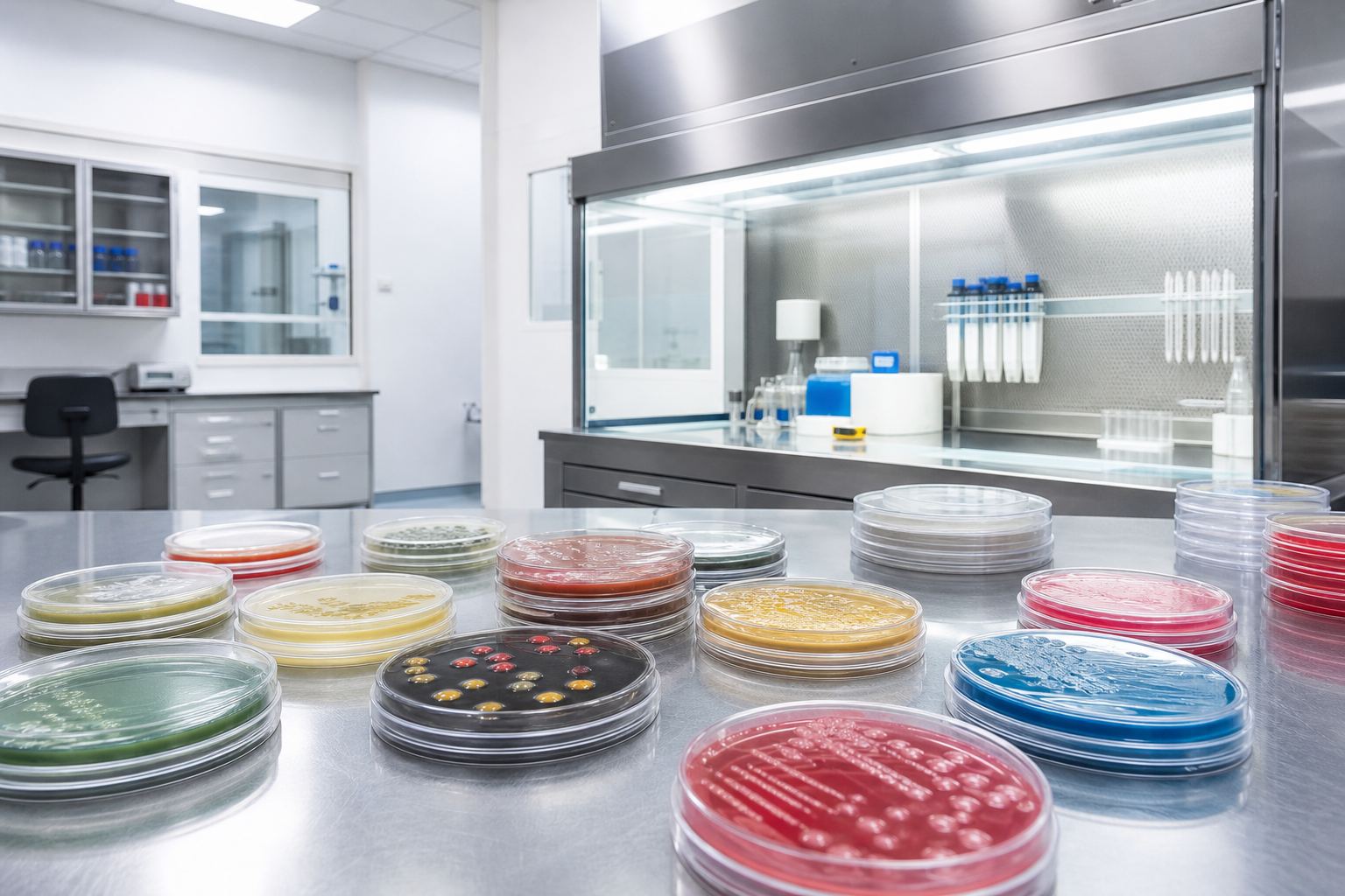
Two discoverersClear spots A cloudy lawn of bacteria on agar, pocked with clear holes. AI-generated still.
bacterio- + phagein, “to devour”d’Hérelle’s word, from Greek
The name
1 plaque ≈ 1 phage
so counting plaques counts phages
Still used today
Chapter I · The bacteria eaters
Twort and d’Hérelle
American Association of Immunologists: in 1915 Frederick Twort described in The Lancet a “glassy and transparent” change on agar plates; the agent passed through a fine porcelain filter that traps bacteria and could be transmitted over many generations; his work was interrupted when he joined the Royal Army Medical Corps. In 1917 Félix d’Hérelle at the Pasteur Institute, studying bacteria from dysentery patients, described an “invisible microbe that is antagonistic to the dysentery bacillus” and named it the bacteriophage, from Greek phagein, to devour. Institut Pasteur: in Tunisia in 1915 he had seen “clear circles” in cultures of a bacterium that attacked locusts.
The 3D scenes are simplified teaching models, not to scale.
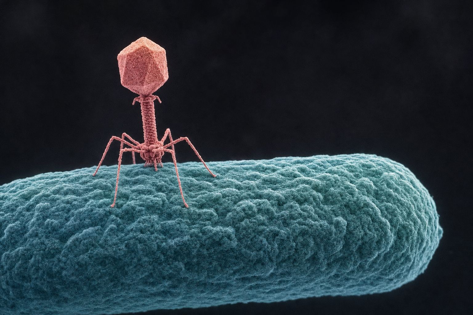
Nobody saw a phage until the electron microscope. The first picture, taken in 1939, showed that many phages look like tiny machines.
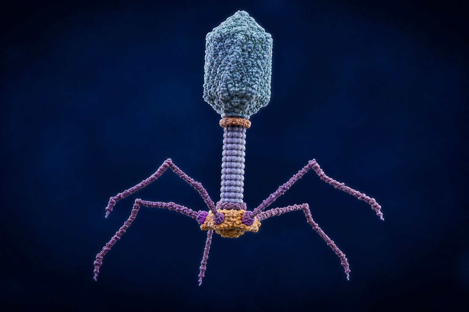
Take T4, a phage that infects the gut bacterium E. coli. Its DNA is packed into a head about 120 nanometres long, more than ten times smaller than the bacterium it infects.
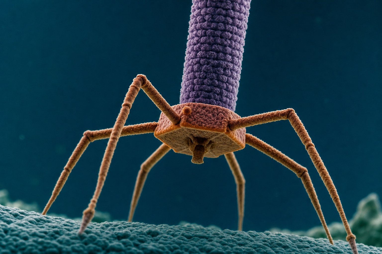
Below the head is a hollow tail, wrapped in a sheath that can contract. At the bottom, a baseplate with six long fibres, like legs.
Press inject, and watch the sheath shorten and drive the tube in.
1939
first electron micrograph of a phage, which showed it was a virus
First lookThe baseplate Close up on the foot of a T4-like phage: the baseplate and its six long fibres. AI-generated still.
120 × 86 nm
the head
~100 nm
the tail
6 + 6
long and short tail fibres
T4 by the numbers (Fokine et al., 2013)
It shrinks to less than half its length
The rigid inner tube is pushed out
DNA leaves through the tube
When the sheath fires
Chapter II · How a phage kills
Anatomy of T4
Fokine et al. (J Mol Biol, 2013): T4 has a 1,200 Å long, 860 Å wide head and a 1,000 Å long tail, with a rigid central tube inside a contractile sheath that shrinks to less than half its length during infection; a multiprotein baseplate at the end of the tail carries six long and six short tail fibres, and the genomic DNA exits through the tail. AAI: the first electron micrograph of a phage was captured in 1939 and published in 1940, showing it was a virus.
Try it: press Inject.
The 3D scenes are simplified teaching models, not to scale.
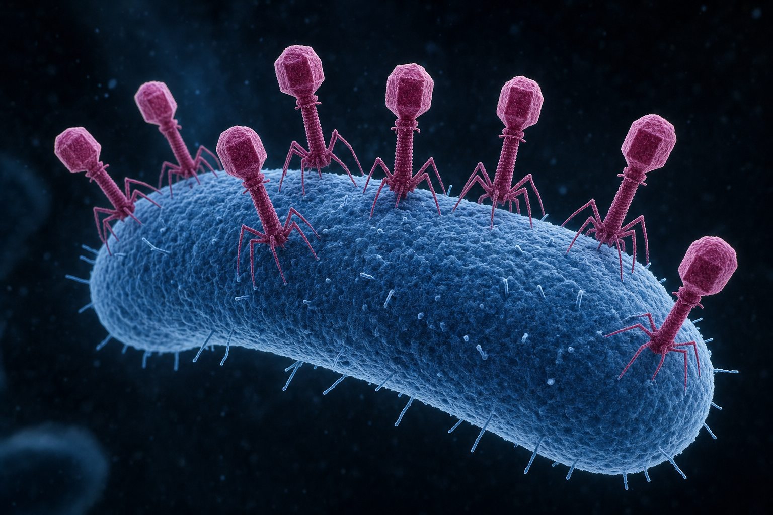
A phage cannot swim or steer; it drifts until it bumps into a cell it can infect.
Its tail fibres grip particular molecules on the cell surface, called receptors. That is why most phages infect only a narrow range of bacteria.
Once it is anchored, the sheath contracts and the tail punches through the cell wall like a syringe.
The DNA slides in. The empty head and tail stay outside, their job done.
Landing Phages meet bacteria by chance, carried by the water around them. AI-generated still.
Lipopolysaccharides on the outer membrane
OmpC, an outer-membrane protein
Receptors T-even phages use (OpenStax)Many at once Several phages can land on one cell, each gripping the surface with its fibres. AI-generated still.
empty coat
head and tail stay on the surface
Left behind
Chapter II · How a phage kills
Attachment and injection
OpenStax Microbiology 6.2: in the lytic cycle of T-even phages the phage attaches to specific receptors on the bacterial surface, such as lipopolysaccharides and the OmpC protein, and most phages have a narrow host range; the tail sheath contracts and acts “like a hypodermic needle to inject the viral genome through the cell wall and membrane”, while the head and other components stay outside.
Try it: replay the landing.
The 3D scenes are simplified teaching models, not to scale.
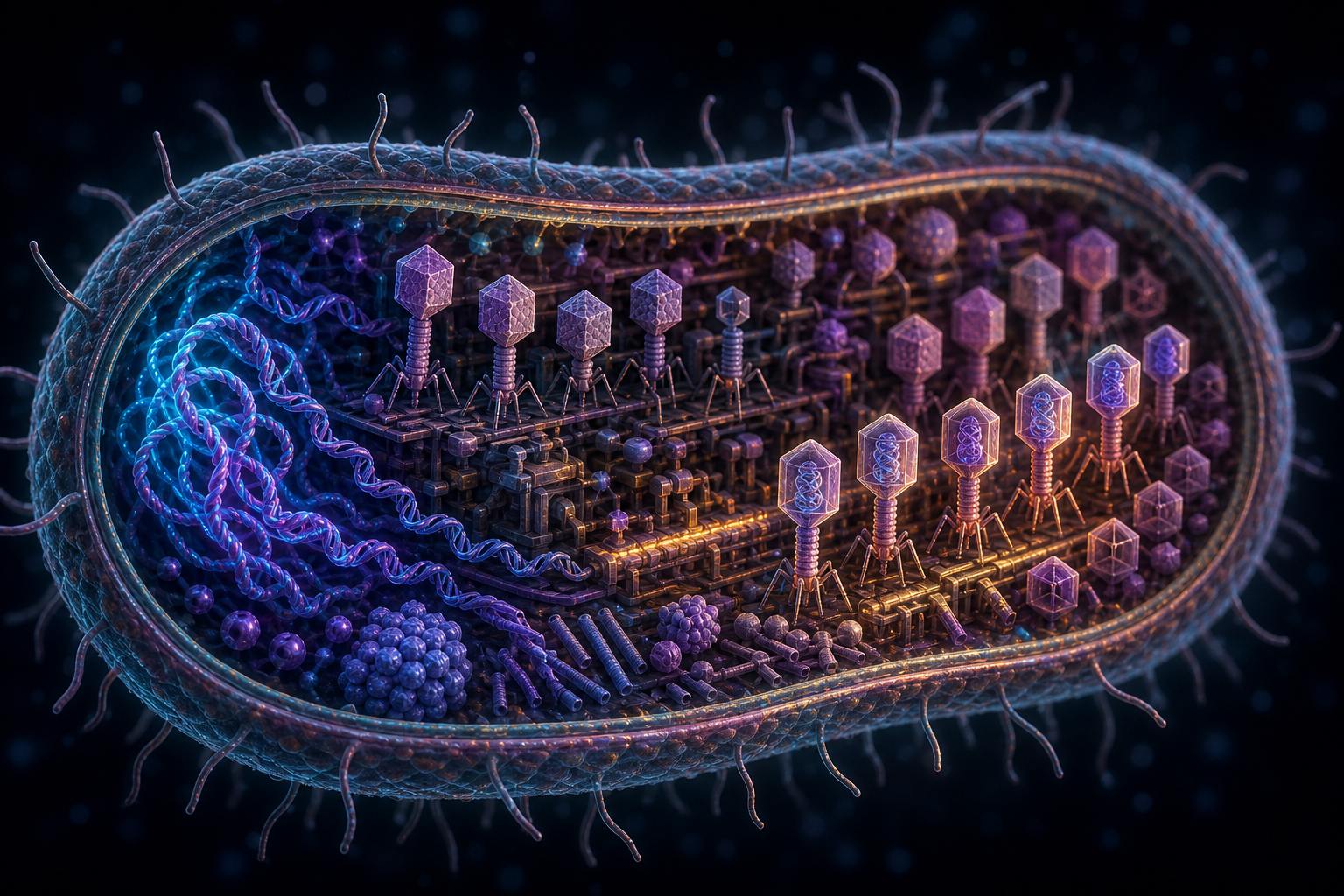
Now the takeover begins. Phage enzymes chop up the bacterium's own DNA, and the cell starts reading phage genes instead.
Early genes make the tools to copy phage DNA. Later genes make heads, tails and fibres, which assemble into new phages.
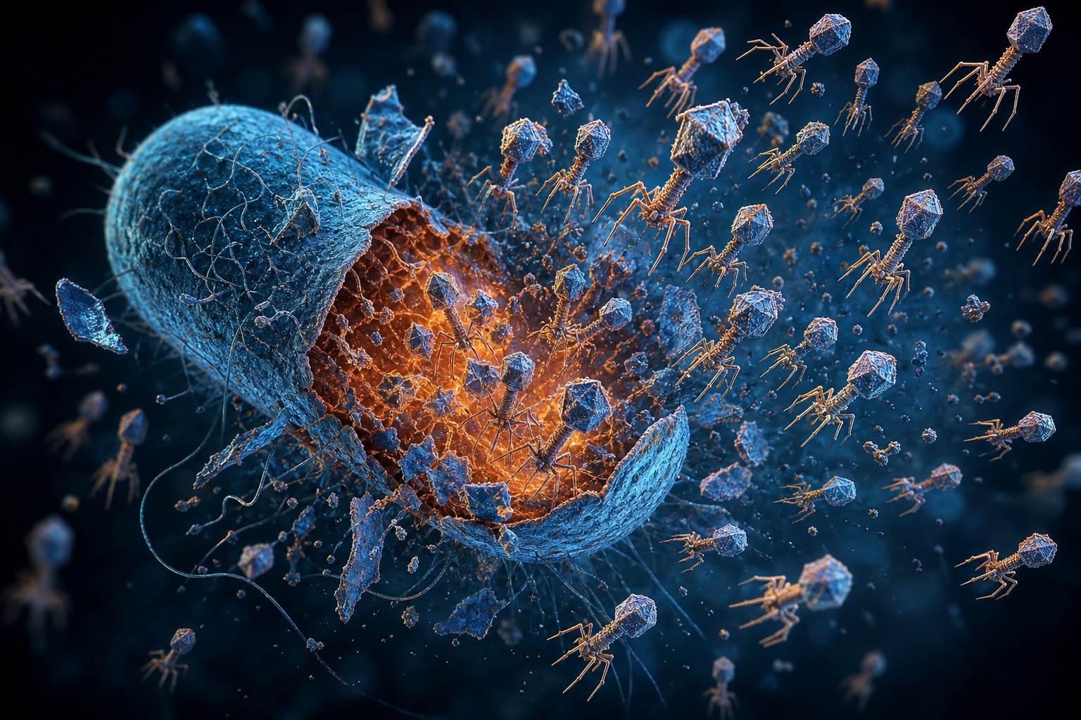
Last come two killers. A protein called holin punches holes in the soft membrane just inside the wall, and an enzyme called endolysin slips through and digests the wall itself.
The cell bursts. In the lab, the classic phages of E. coli release about 120 to 300 new phages. This is the lytic cycle.
1 Attach
2 Inject
3 Hijack and copy
4 Assemble
5 Burst
The lytic cycleA virus factory Heads are filled with DNA before tails are attached. AI-generated still.
Holin: timed holes in the inner membrane
Endolysin: digests the peptidoglycan wall
The two killersLysis Burst sizes for the T phages of E. coli B are 120 to 300, by classic counts. AI-generated still.
Chapter II · How a phage kills
Hijack, assemble, burst
OpenStax 6.2: virus-encoded endonucleases degrade the bacterial chromosome; genes for copying the genome are expressed early and those for capsid and tail proteins later; new virions assemble and are released when phage proteins such as holin or lysozyme break the cell wall. Wikipedia (Lytic cycle): holin accumulates in the inner membrane until it suddenly forms holes, letting the endolysin reach and attack the peptidoglycan; for lambda, lysis comes about 50 minutes after infection with about 100 virions. BioNumbers 105870: the T-system phages of E. coli B release 120 to 300 virions per burst.
Try it: replay the cycle.
The 3D scenes are simplified teaching models, not to scale.
One burst becomes a hundred phages. Each finds a neighbour, and a hundred bursts become ten thousand.
That is how a single phage clears a hole in a lawn of bacteria big enough to see with your eye.
Release a phage onto the lawn, and change the burst size to see how fast the plaque grows.
start
1
round 1
100
round 2
10,000
round 3
1,000,000
Phages after each round, burst of 100
Seen by eye One plaque, a few millimetres across, on a plate held up to the light; the lawn around it is millions of cells. AI-generated still.
Each infected cell bursts after a fixed delay
Phages reach only nearby cells
A bigger burst infects more neighbours
In the model
Chapter II · How a phage kills
Why one phage makes a hole you can see
Each round of infection multiplies the number of phages by the burst size, so growth is exponential: with 100 per burst, three rounds turn one phage into a million. On a plate, phages spread only to neighbouring cells, so the cleared zone grows outward into a plaque, one plaque per original phage, which is how phages are counted. The lawn on screen is a simplified model, not a simulation of real diffusion.
Try it: release a phage, then try a burst of 50, 150 or 300.
The 3D scenes are simplified teaching models, not to scale.
Not every phage kills right away. Some can wait. They are called temperate phages, temperate meaning mild, and the best studied is phage lambda.
Its DNA stitches itself into the bacterium's chromosome and stays there, silent. In that state it is called a prophage.
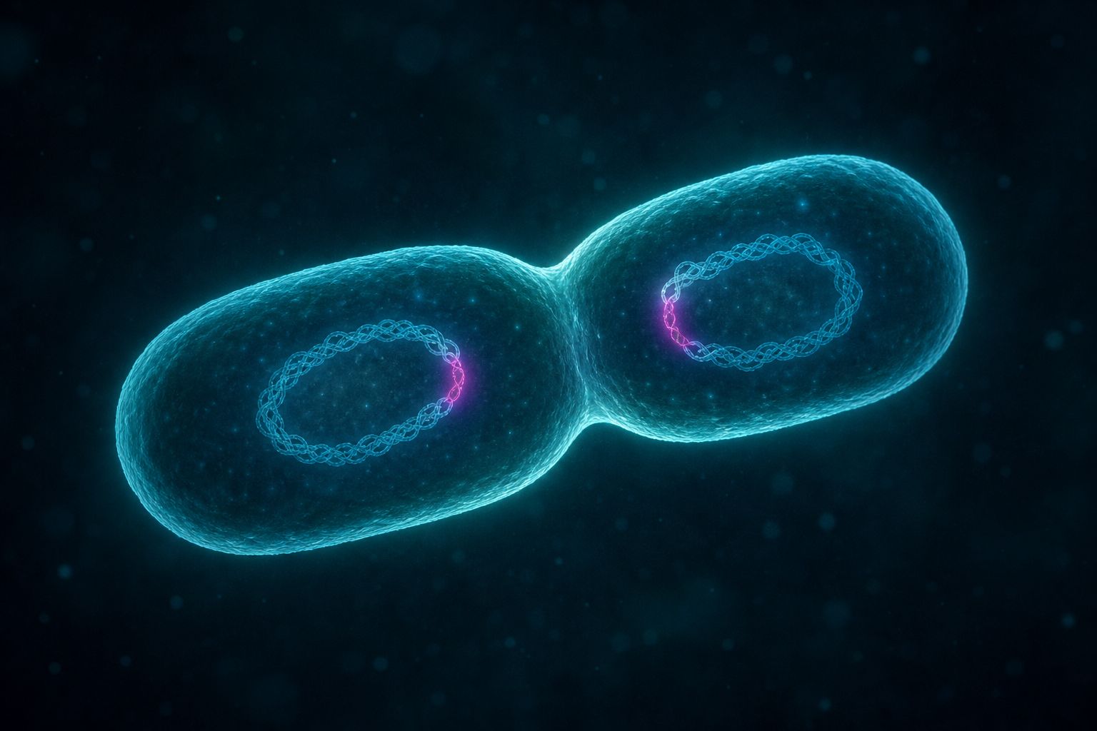
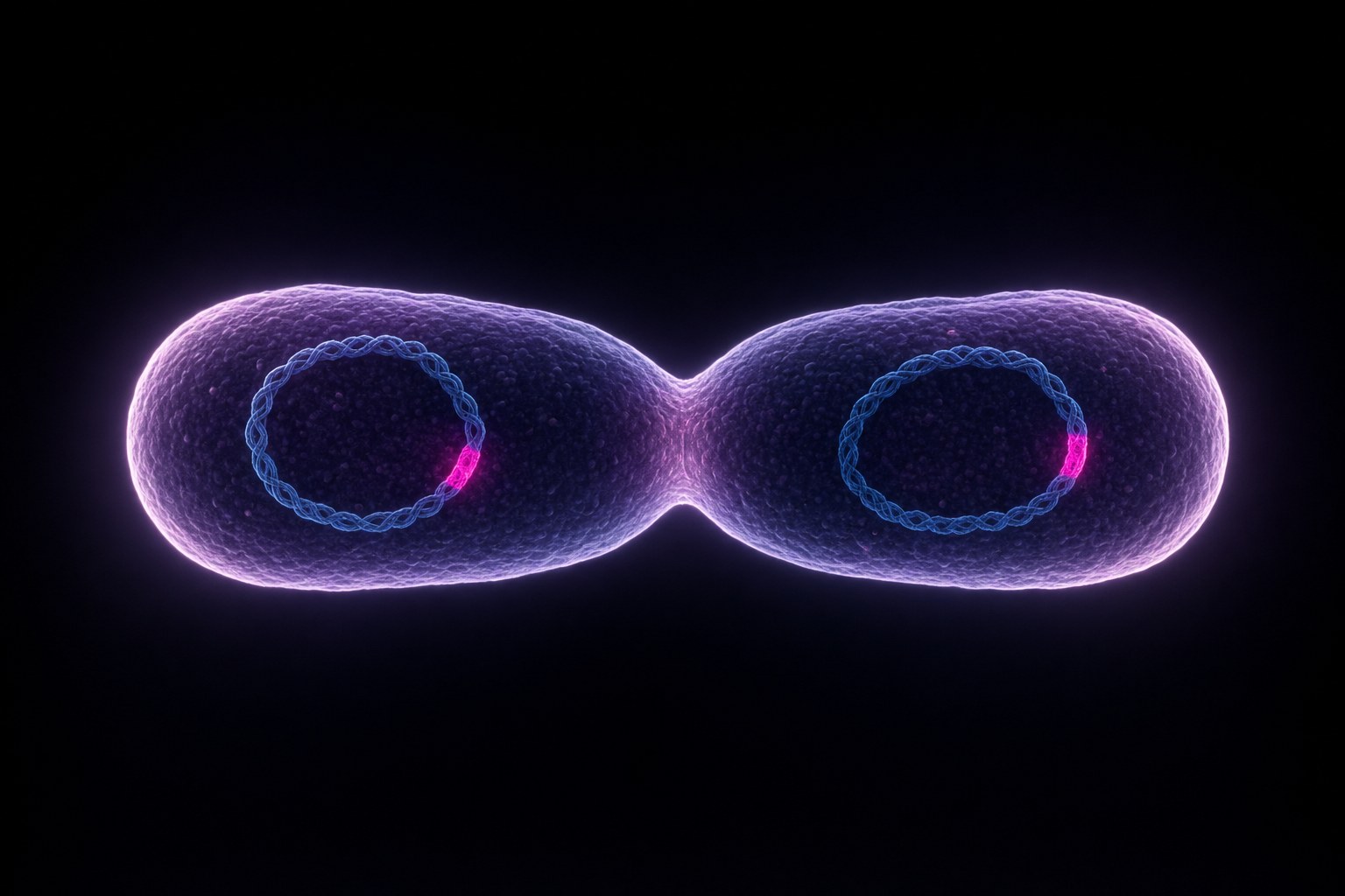
Every time the bacterium divides, it copies the prophage along with its own genes. This is the lysogenic cycle.
But danger to the cell, such as ultraviolet light or harmful chemicals, can wake it. It cuts itself out and turns lytic. Try both paths.
Virulent: always lytic, such as T4
Temperate: lytic or lysogenic, such as lambda
Two kinds of phageA hidden genome The magenta segment is the viral DNA, carried along at every division. AI-generated still.
prophage
the phage DNA inside the chromosome
lysogen
a bacterium that carries one
Words to know
Ultraviolet light
Chemical exposure
Starvation
What can wake it (OpenStax)
Chapter III · The quiet passenger
The lysogenic cycle
OpenStax 6.2: temperate phages such as lambda can enter the lysogenic cycle, in which the phage genome integrates into the bacterial chromosome as a prophage; a bacterium carrying a prophage is a lysogen, and the prophage is copied and passed to daughter cells. Ultraviolet light, chemical exposure or starvation can induce the prophage to excise and enter the lytic cycle.
Try it: lysogenic, lytic, or a flash of UV.
The 3D scenes are simplified teaching models, not to scale.
A quiet passenger can change the bacterium it lives in: some prophages carry genes the bacterium then uses.
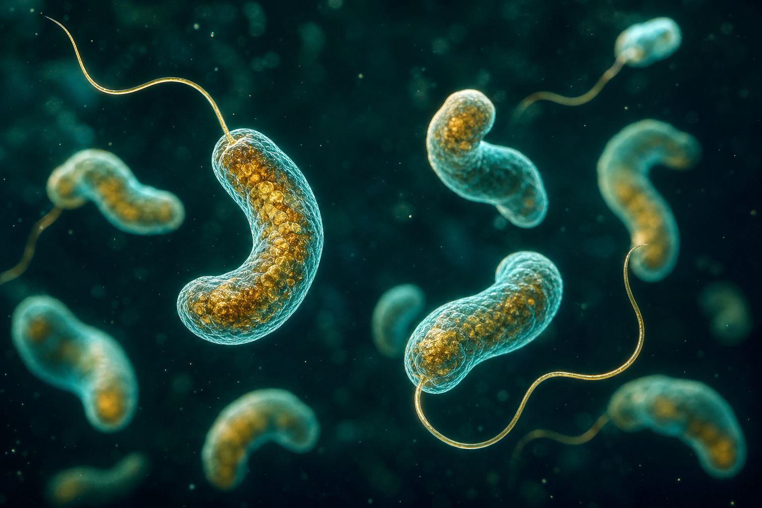
The toxin that makes cholera so dangerous is encoded by a phage, and so are some forms of the botulinum toxin. This is called lysogenic conversion: the passenger converts its host.
Phages also carry stray pieces of bacterial DNA from one cell to the next, which helps bacteria evolve.
Vibrio cholerae Comma-shaped cholera bacteria, each with one whip-like tail. AI-generated still.
Vibrio cholerae: cholera toxin, severe diarrhoea
Clostridium botulinum: some forms of botulinum toxin, which can paralyse
Toxins carried by phages (OpenStax)
Generalised: a random piece, packed by mistake
Specialised: the genes next to the prophage
Transduction, two kindsPassed on A dividing bacterium: both daughter cells inherit the prophage, drawn here as a bright band in the DNA, and whatever genes it brought. AI-generated still.
Chapter III · The quiet passenger
Lysogenic conversion and transduction
OpenStax 6.2: a prophage can change the traits of its host, called lysogenic or phage conversion; the toxin of Vibrio cholerae, which can cause severe diarrhoea, and that of Clostridium botulinum, which can cause paralysis, are given as phage-encoded examples. Only some forms of botulinum toxin are carried by phages; in other strains the toxin genes sit on the chromosome or on a plasmid, so the film says “some forms”. In transduction a phage transfers bacterial DNA between cells: generalised transduction moves a random piece packaged by mistake in the lytic cycle, specialised transduction moves DNA next to the integration site; transduction may help drive bacterial evolution.
This is biology education, not medical advice.
The 3D scenes are simplified teaching models, not to scale.
Phages also answered one of biology's biggest questions: what are genes made of, protein or DNA?
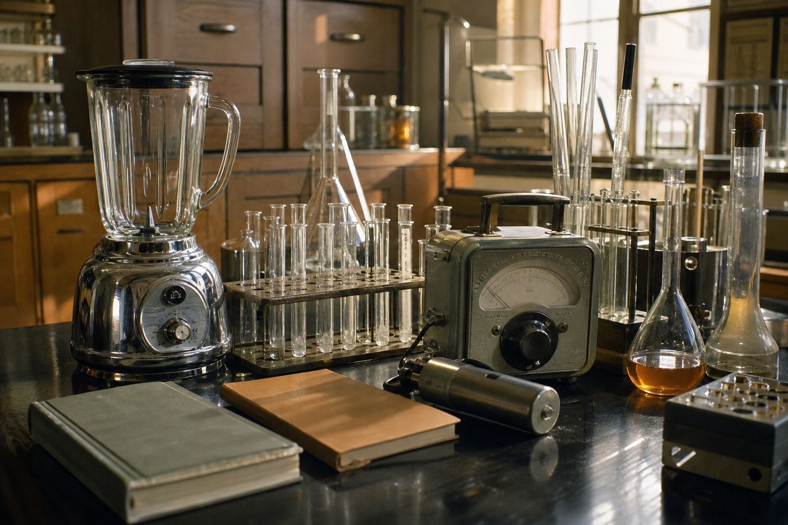
In 1952, at Cold Spring Harbor in New York, Alfred Hershey and Martha Chase grew two batches of phages: one labelled with radioactive phosphorus, which marks DNA, the other with sulfur, which marks protein.
They let the phages infect bacteria, then shook the empty coats off in a kitchen blender.
The DNA label had gone into the cells. The protein label had mostly stayed outside. So the phage's genes were in its DNA, not its protein. Label each one and run the blender.
A phage is just protein and DNA
Only one of them enters the cell
So that one must carry the genes
The question
³²P
phosphorus: in DNA, not in protein
³⁵S
sulfur: in protein, not in DNA
Two labelsA kitchen tool A blender, centrifuge tubes and a radiation counter: the kit of a famous result. AI-generated still.
Hershey
shared it with Max Delbrück and Salvador Luria
Nobel Prize, 1969
Chapter IV · What phages taught us
Hershey and Chase, 1952
Cold Spring Harbor Laboratory: in 1952 Alfred Hershey and Martha Chase used bacteriophages labelled with radioactive phosphorus-32 in their DNA and sulfur-35 in their protein; a Waring kitchen blender separated the viral material from the bacteria, and the result showed that DNA, not protein, carries the genetic information. Hershey shared the 1969 Nobel Prize in Physiology or Medicine with Max Delbrück and Salvador Luria.
Try it: label the DNA or the protein, then run the blender.
The 3D scenes are simplified teaching models, not to scale.
Bacteria are not helpless. They fight back, and many of them keep a memory of past infections, written in their own DNA, in a region called CRISPR.
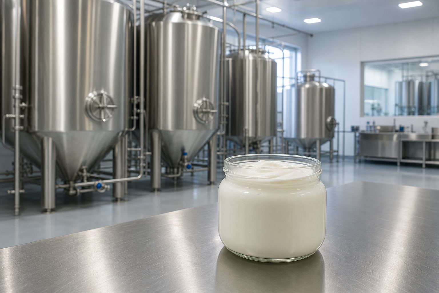
In 2007 scientists at the food company Danisco, working with yogurt bacteria, showed what it does.
Bacteria that survived an attack had added a short piece of that phage's DNA, a spacer, to their CRISPR region. After that they resisted the same phage.
Remove the spacer, and the protection was gone. Press attack twice: the first phage gets in and leaves a spacer behind; the second is cut to pieces.
repeat · spacer · repeat
short repeats with stored phage snippets between them
CRISPRYogurt cultures Phages that kill starter bacteria can ruin a batch, so dairy firms study them closely. The bacterium here was Streptococcus thermophilus. AI-generated still.
Survivors gained new spacers
Each resisted the phage that matched
Adding a spacer gave resistance
Barrangou et al., Science, 2007Memory as DNA A guide strand lines up with matching DNA before it is cut. AI-generated still.
Chapter IV · What phages taught us
CRISPR, the bacterial immune memory
Embryo Project (Arizona State University): Rodolphe Barrangou, Philippe Horvath and colleagues at Danisco reported in Science (2007) “CRISPR Provides Acquired Resistance against Viruses in Prokaryotes”, working with Streptococcus thermophilus, a yogurt culture bacterium; strains with spacers matching a virus resisted it, removing the spacer made them vulnerable, adding a matching spacer gave resistance, and survivors of infection had gained new spacers; inactivating the cas5 gene removed resistance.
Try it: attack twice. The first attack stores a spacer; the second is blocked.
The 3D scenes are simplified teaching models, not to scale.
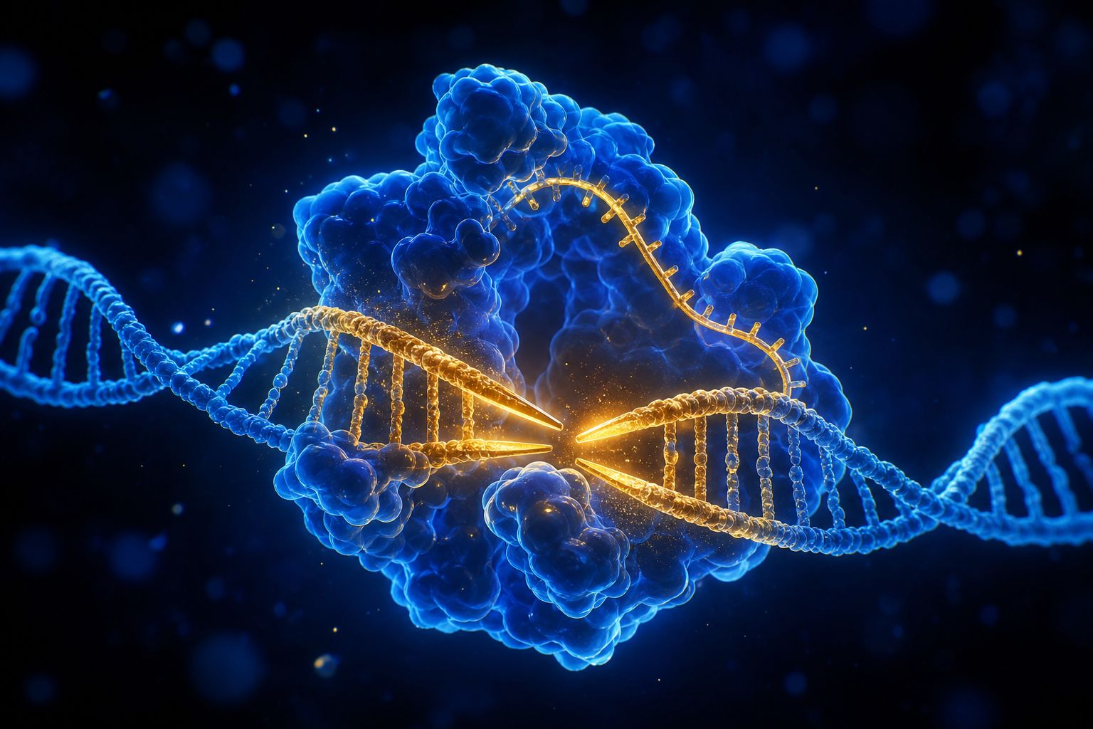
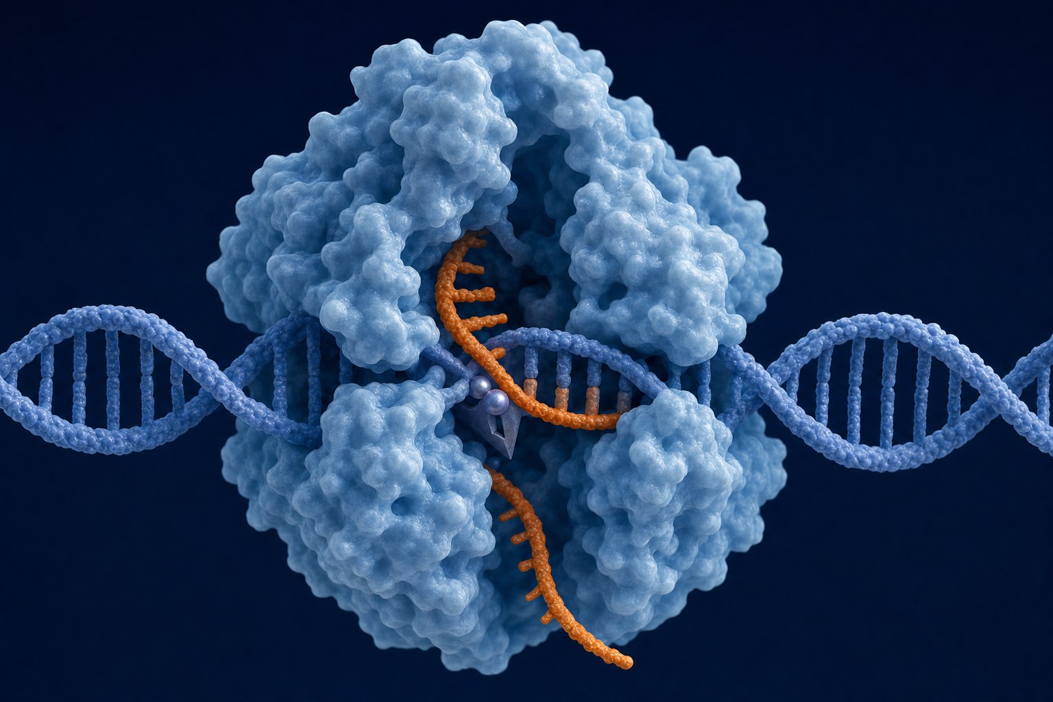
Matching proteins called Cas use the stored spacer to find the invader's DNA, and cut it.
In 2012 Emmanuelle Charpentier and Jennifer Doudna turned one such system, CRISPR-Cas9, into genetic scissors that cut DNA where scientists choose.
It won them the 2020 Nobel Prize in Chemistry. Labs use it to switch genes off and see what they do, and to work on crops and on treatments for inherited disease. A tool that rewrites genes began as a bacterium's defence against phages.
Search and cut A Cas protein, holding a short RNA guide copied from a spacer, closes on the matching DNA. AI-generated still.
2007 CRISPR shown to defend against phages
2011 Charpentier finds tracrRNA in Streptococcus pyogenes
2012 CRISPR-Cas9 genetic scissors
2020 Nobel Prize in Chemistry
From yogurt to genome editingIn labs everywhere Gene editing is now routine research work. AI-generated still.
“for developing a method of genome editing”the Nobel citation, as UCMR quotes it
Umeå University, on the 2020 prize
Chapter IV · What phages taught us
Genetic scissors
Umeå University (UCMR): Emmanuelle Charpentier and Jennifer Doudna received the 2020 Nobel Prize in Chemistry for “developing a method of genome editing”. While studying Streptococcus pyogenes, Charpentier discovered a new molecule, tracrRNA, and published it in Nature in 2011, the year she began collaborating with Doudna; in 2012 the two showed in Science how Cas9, guided by two RNAs, can be aimed at a chosen gene, and the tool has since been adopted by researchers around the world.
Try it: attack a bacterium that already remembers.
The 3D scenes are simplified teaching models, not to scale.
Phages may be the most numerous biological entities on Earth: microbiologists estimate about ten to the power of thirty-one of them, and the virologist Curtis Suttle reckons they kill up to forty percent of the ocean's bacteria every day.
Nearly all of them carry DNA, so the gut virus in the news matters: the picobirnavirus has RNA genes, which would make it only the third known group of RNA phages, and its discoverers caution that finding it in patients does not prove it causes disease. And if phages kill bacteria on that scale, could we aim them at the ones that make us ill?
~10³¹
phages on Earth, about ten for every bacterium
~50 million
viruses in a millilitre of seawater
up to 40%
of ocean bacteria killed each day
Estimates (Science News; Curtis Suttle)The sea Sunlit water teeming with microbes and the viruses that prey on them. AI-generated still.The gut Gut virus counts run to 10⁹–10¹⁰ particles per gram of stool, and most gut phages are thought to live quietly as prophages (Frontiers, 2024). AI-generated still.
Nearly all phages: DNA genes
RNA phages: thought to be rare
Picobirnaviruses: proposed as the third group (2026)
RNA phages
Chapter V · A planet of phages
A planet of phages
Science News: microbiologists estimate about 10³¹ bacteriophages worldwide, consistent with about 10 phages per bacterium and some 10³⁰ bacteria; Curtis Suttle estimates about 50 million viruses per millilitre of seawater and that phages destroy up to 40 percent of the bacteria in Earth’s oceans each day. BioNumbers (after Minot et al., PNAS 2013): at least 10⁹ virus-like particles per gram of human feces. Yu et al. (Frontiers in Microbiology, 2024): roughly 10⁹ to 10¹⁰ particles per gram, and “the majority of phages found in the human gut are prophages.” WashU (2026): the new study calls Picobirnaviridae the third clade of RNA bacteriophages.
These are estimates, not counts, and they are often quoted with wide uncertainty.
The 3D scenes are simplified teaching models, not to scale.
D'Hérelle saw the medical promise at once: a living cure that hunts the germ and leaves the patient alone.
In 1923 the microbiologist George Eliava opened an institute in Tbilisi, Georgia, where d'Hérelle later worked. Phage drops and pills were sold in pharmacies in the Soviet Union and later across Eastern Europe.
In the West, sulfa drugs and then antibiotics arrived, and phage therapy was set aside: they killed more kinds of bacteria, were easier to make, and a 1934 review in JAMA doubted phages worked inside the body.
But bacteria evolve resistance. The World Health Organization says drug-resistant infections directly caused about 1.27 million deaths in 2019.
He suggested they could be used to treat bacterial infections.of Félix d’Hérelle
Institut Pasteur, on 1917A phage institute Shelves of phage preparations in a 1930s laboratory. Eliava was arrested and executed by the Soviet authorities in 1937. AI-generated still.At the pharmacy Phage ointments, pills and drops were sold cheaply across Eastern Europe. AI-generated still.
Sulfa drugs were cheaper and easier to make
They covered more kinds of infection
A 1934 JAMA review doubted phages worked in the body
Why the West set phages aside (AAI)
1.27 million
deaths caused directly by drug resistance
4.95 million
deaths associated with it
WHO, 2019 figures
Chapter VI · Phages as medicine
A cure set aside, and found again
Institut Pasteur: in 1917 d’Hérelle suggested phages could treat bacterial infections; after the war they were supplanted by antibiotics. Wikipedia (George Eliava Institute): the institute opened in Tbilisi in 1923; d’Hérelle worked there for about 18 months after 1934; Eliava was executed in 1937; the institute became the Soviet centre for phage drugs, and phage ointments, pills, drops and rinses were sold cheaply in pharmacies across Eastern Europe. AAI: sulfa drugs in the 1930s were cheaper, easier to produce and covered more infections, and a 1934 JAMA review doubted phages worked in the body. WHO (2023): antimicrobial resistance was responsible for about 1.27 million deaths in 2019 and associated with 4.95 million.
This is history and biology education, not medical advice. Talk to a doctor about any infection.
The 3D scenes are simplified teaching models, not to scale.
So phages are being looked at again. Their pickiness is both their strength and their difficulty: a phage kills only the strains it recognises.
Release one on a mixed lawn and see which bacteria it spares.
In the United States no phage medicine is approved; doctors use them case by case, with special permission, when nothing else works.
One programme in Pittsburgh reported encouraging results at a conference in 2026, warning that phages are not a miracle cure and that proper clinical trials are what is needed.
Spares the body’s helpful bacteria
Must be matched to each infection
Bacteria can evolve resistance to phages too
Pickiness, both waysMatching the phage A patient’s bacteria are grown and tested against a library of phages. AI-generated still.
Healio (26 October 2026), reporting a conference presentation by Ghady Haidar (University of Pittsburgh) at IDWeek: the Pittsburgh Phage Program received 357 requests from November 2019 to April 2026; phages were found for 70.9% of isolates screened; among 26 patients with adjudicated day-28 outcomes, 41% improved and 36% were stable; clearance was often followed by relapse; no deaths were attributed to phages. Haidar said there are no FDA-approved phage products in the US, that phages are used through single-patient applications and compassionate use, that they are “not a miracle cure”, and that the field must move to clinical trials. OpenStax 6.2: most phages have a narrow host range.
A conference report, not yet a peer-reviewed trial. This is education, not medical advice; talk to a doctor.
Try it: release the phage on the mixed lawn.
The 3D scenes are simplified teaching models, not to scale.
Phages showed us that DNA, not protein, carries genes, gave us genetic scissors, and still shape the oceans and our own gut.
And in your gut right now, trillions of bacteria carry prophages that may never wake. What are those quiet passengers doing to them, and to you? A century after Twort and d'Hérelle, nobody can say.
1915–1917 Twort and d’Hérelle discover phages
1923 Eliava’s institute opens in Tbilisi
1939 first electron micrograph of a phage
1952 Hershey–Chase: DNA, not protein, carries genes
2007 CRISPR shown to fight phages
2020 Nobel Prize for CRISPR gene editing
2026 a common gut virus proposed as a phage
The bacteria eaters: a timelineStill hunting Phages descending on a bacterium. AI-generated still.
What do quiet prophages do to their hosts, and to us?
How many gut viruses are really phages?
Can phage therapy pass proper clinical trials?
Open questions
Epilogue · Sources
Sources
Every fact in this film comes from the sources below. The chapter stills and pictures are AI-generated artist’s impressions; they show no real microbes, people or places. The phage, the infected cell, the lawn of bacteria, the blender experiment and the CRISPR scene in 3D are simplified teaching models, not to scale. The 2026 picobirnavirus study is a single new result, reported here as its authors and their university describe it. Numbers such as 10³¹ phages are estimates. Nothing here is medical advice.