PlanetPast

1986 → 2026 · Neuroscience meets AI

The Fly in the Machine

How scientists and AI mapped every neuron of a fruit fly, how the open-source wiring diagram became a running brain model and a virtual body, and what the viral 'fly plays Doom' demos really show about brains and artificial intelligence.

Interactive 3D film7 min1986 to 2026Published 2026-10-13

Watch the interactive filmอ่านภาษาไทย

Prologue

September 2026 · An open-source brain

The fly in the machine

Every neuron and every connection of a fruit fly’s central nervous system, free for anyone to download.

In September 2026, scientists in Britain and the United States, working with Google, released a complete wiring map of a male fruit fly’s nervous system.

It traces about 166,700 neurons and well over a hundred million connections between them. And it is open: anyone can download it.

Within days, people had wired the map into Minecraft, Doom and other games, and the videos went viral.

What exactly was mapped? How do you map a brain at all? And is this really a brain running inside a computer?

Prologue · Why the news matters

The fly in the machine

On 3 September 2026 a team from the MRC Laboratory of Molecular Biology and the University of Cambridge, HHMI’s Janelia Research Campus and Google Research published in Cell the first complete connectome (wiring diagram) of an adult male fruit fly’s central nervous system: its brain, both optic lobes and its ventral nerve cord. The data are open for anyone to use.

Neurons
166,700
brain, optic lobes and nerve cord
Synaptic connections
~124 million
as reported by UKRI / MRC
Neuron types
~11,700
classified by the team
Imaging
7 electron microscopes, one year
the nervous system was cut into 66 pieces

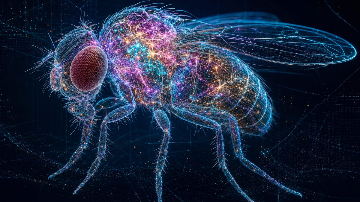
The fly in the scene is a simplified model; the glowing points inside its head and thorax stand for its brain and nerve cord.

How to use: pick a chapter below the scene or press ← → to change step · ▶ plays the step · drag to rotate, scroll or pinch to zoom · buttons in the box at the top right let you try things yourself · “Play the film” runs every step full-screen.

Sources: UKRI / MRC LMB: world-first map of a male fly brain (Sept 2026); Google: the male fruit fly brain map (Sept 2026); Live Science: map of a male fly central nervous system (Sept 2026); 36Kr: MaleCNS and the Minecraft demo

Chapter I · Wiring diagrams

Chapter I · The idea

A map of who talks to whom

A connectome lists every neuron and every connection between them.

A brain is a network. Its cells, the neurons, pass signals to one another across tiny junctions called synapses.

A connectome is the complete list: every neuron, and every synapse that links one neuron to another.

Think of it as the wiring diagram of a circuit board, or a map of every road in a country.

A map shows which routes exist. It does not show the traffic. That difference will matter later.

Chapter I · Step 1

A map of who talks to whom

A connectome is a complete wiring diagram of a nervous system. Each neuron is a node; each synapse is a link from one neuron to the next. Counting how many synapses join two neurons gives a rough idea of how strongly they are connected.

  1. What it shows: which neurons connect, in which direction, and through how many synapses.
  2. What it leaves out: how strong each synapse really is, how it changes with learning, and the chemistry (hormones, neuromodulators) that tunes the whole network.

Press “Send a signal” to watch activity spread through the small network in the scene.

Sources: University of Cambridge: first map of every neuron in an adult fly brain (2024); Stork.ai: the viral demos explained

Chapter I · 1986

The first wiring diagram

A worm with 302 neurons, traced by hand from electron micrographs.

The first connectome belonged to a tiny worm, Caenorhabditis elegans, about a millimetre long.

Its adult hermaphrodite has just 302 neurons. A team in Cambridge, led by John White and Sydney Brenner, traced them through thin slices seen with an electron microscope.

The work was done largely by hand, and the full map was published in 1986.

For decades, it was the only complete animal connectome. Doing the same for an insect brain looked impossible.

Chapter I · Step 2 · 1986

The first wiring diagram

In 1986 John White, Eileen Southgate, Nichol Thomson and Sydney Brenner published “The structure of the nervous system of the nematode Caenorhabditis elegans” in the Philosophical Transactions of the Royal Society. It named and mapped all 302 neurons of the adult hermaphrodite worm, reconstructed from electron micrographs of serial sections.

Worm (1986) 302 Fly brain (2024) 139,255 Fly CNS (2026) 166,700

The male fly’s nervous system has about 550 times as many neurons as the worm. Bars are to scale.

Sources: OpenWorm: White et al. 1986; 36Kr: MaleCNS and the Minecraft demo

Chapter II · Mapping a brain

Chapter II · Slice and image

Thinner than a soap bubble

To see every wire, the brain is cut into thousands of slices and photographed with electrons.

A fly brain is about the size of a poppy seed. Its finest wires are far too thin to see with ordinary light.

So the brain is preserved, stained with heavy metals, and either sliced or shaved away, layer by layer.

For the female fly brain mapped in 2024, about seven thousand slices, each forty nanometres thick, were imaged with electron microscopes at Janelia.

That produced more than a hundred terabytes of images. For the male fly, seven microscopes ran for a year.

Chapter II · Step 3

Thinner than a soap bubble

Neurons’ finest branches are only tens of nanometres wide, below the resolution of light microscopes. Electron microscopes can see them, but only in very thin layers. So the brain is cut into a stack of images, like a deck of cards, and the 3D shapes are rebuilt from the stack.

Female fly brain (FlyWire, 2024)
~7,000 slices
each about 40 nm thick
Image data
> 100 TB
for one brain
Male fly CNS (2026)
Seven electron microscopes running for a year
surface imaged, a thin layer shaved off, repeat; the tissue was first cut into 66 pieces

In the scene, slices come off the block and stack up to the right. Each slice is drawn far thicker than in reality.

Sources: University of Cambridge: first map of every neuron in an adult fly brain (2024); UKRI / MRC LMB: world-first map of a male fly brain (Sept 2026)

Chapter II · 2018

AI follows the wires

A neural network colours in each neuron, slice after slice.

Tracing every neuron by hand through millions of images would take a human team many centuries.

So the tracing is done by AI. In 2018 Google researchers described flood-filling networks: start at one point inside a neuron, and let a neural network fill outward until it hits the cell’s edge.

Repeated slice after slice, each colour becomes one cell in 3D.

For the male fly, Google’s models segmented the tissue; the team reports more than a thousandfold saving in time and cost.

Chapter II · Step 4 · 2018

AI follows the wires

In a flood-filling network, a recurrent convolutional neural network starts from a seed point inside one neuron and repeatedly predicts which nearby pixels belong to the same cell, “filling” it until it reaches the membrane. Google researchers reported in 2018 that this improved the accuracy of automated reconstruction by about an order of magnitude.

  1. Segment: AI splits each image into regions, one per cell cross-section.
  2. Link: regions are joined across slices into 3D neurons.
  3. Find synapses: other models spot the contacts where one neuron signals to another.

FlyWire’s AI for the female brain was developed at Princeton; for the male nervous system, Google Research built the segmentation models with Janelia. Press “Seed a neuron” to restart the fill.

Sources: Google Research: flood-filling networks (2018); University of Cambridge: first map of every neuron in an adult fly brain (2024); Neuroscience News: microscopy and AI decipher the fly’s neural architecture

Chapter II · Proofreading

People fix the AI’s mistakes

Automatic tracing still merges some neurons and cuts others in two.

The AI is fast, but not perfect. It sometimes joins two neurons into one, or breaks one neuron into pieces.

So people proofread. For the female brain, the FlyWire consortium, with more than 280 researchers and volunteers from the public, spent an estimated 33 person-years fixing errors.

Experts then named the cells and sorted them into types.

The result is not a picture of a brain. It is a database you can query, one neuron at a time.

Chapter II · Step 5

People fix the AI’s mistakes

Automatic segmentation makes two kinds of error: merges (two neurons glued into one) and splits (one neuron broken into fragments). Because a single wrong join can create thousands of false connections, every neuron is checked.

FlyWire proofreading
~33 person-years
female brain, 2024
FlyWire consortium
76+ labs
287 researchers, plus members of the public

In the scene, one reconstructed neuron has been wrongly merged with its neighbour (flashing red). Press “Fix the error” to split it.

Sources: University of Cambridge: first map of every neuron in an adult fly brain (2024); UKRI / MRC LMB: world-first map of a male fly brain (Sept 2026)

Chapter III · The fly maps

Chapter III · 2020 → 2026

From half a brain to a whole nervous system

Each map was bigger, more complete, and open to everyone.

In 2020, Janelia and Google released the hemibrain: about 25,000 neurons from the central part of a fly brain.

In October 2024, the FlyWire consortium published the first map of an entire adult fly brain: 139,255 neurons and about fifty million connections, from a female fly.

In 2026 came the male: brain, optic lobes and the ventral nerve cord, the insect’s spinal cord, together. About 166,700 neurons.

For the first time, you can follow a signal from the eyes and taste sensors all the way to the muscles of the legs and wings.

Chapter III · Step 6

From half a brain to a whole nervous system

  • 2020 Hemibrain (Janelia and Google): about 25,000 neurons across part of the central brain.
  • 2024 FlyWire (Princeton-led consortium, two papers in Nature): the whole brain of a female fly, 139,255 neurons, about 50 million connections, more than 8,000 cell types.
  • 2026 Male CNS (MRC LMB and Cambridge, Janelia, Google Research; Cell): brain, optic lobes and ventral nerve cord of a male fly, 166,700 neurons, about 11,700 neuron types.

The ventral nerve cord sits in the fly’s thorax and runs the legs and wings, like our spinal cord. Including it links sensing to movement in one map. Use the buttons to compare the three maps.

Sources: Neuroscience News: microscopy and AI decipher the fly’s neural architecture; University of Cambridge: first map of every neuron in an adult fly brain (2024); UKRI / MRC LMB: world-first map of a male fly brain (Sept 2026); Live Science: map of a male fly central nervous system (Sept 2026)

Chapter III · Two sexes

Mostly the same, with switches

With a male and a female map, scientists can compare the wiring cell type by cell type.

With both maps, researchers could compare male and female nervous systems type by type.

About 95 percent of the cells are shared. Most circuits for seeing, tasting and moving are the same.

But some cell types exist only in males, some only in females, and others differ in shape.

A male-specific network appears to coordinate courtship and aggression: the same basic wiring, with switches that route signals differently.

Chapter III · Step 7

Mostly the same, with switches

Comparing the male map with the female FlyWire brain, the team matched neuron types between the sexes.

Same in both sexes
8,069
types matched one to one
Different in shape
138
“dimorphic” types
Male-specific
289
types
Female-specific
71
types

Companion studies described a male-specific network for courtship and aggression, and taste circuits from receptors on the legs, wings and mouth. The authors call the diagram a tool for generating hypotheses: it suggests experiments rather than replacing them. The magenta points in the scene mark sex-specific cells (positions illustrative).

Sources: Neuroscience News: microscopy and AI decipher the fly’s neural architecture; Live Science: map of a male fly central nervous system (Sept 2026); UKRI / MRC LMB: world-first map of a male fly brain (Sept 2026)

Chapter IV · Running the map

Chapter IV · 2024

Switching on the map

Turn every neuron into a simple equation, wire them up as the map says, and press play.

A map is still. To make it run, researchers turn every neuron into a simple equation: add up incoming signals, and fire when a threshold is crossed.

In 2024, Philip Shiu, Kristin Scott and colleagues at Berkeley did this for all 139,255 neurons of the FlyWire brain. It runs on a laptop.

Stimulate the sugar-sensing neurons, and the model predicts which cells fire, all the way to the neurons that drive feeding.

They then tested many of those predictions in real flies, using light to switch neurons on. Many held up. The model is crude, but the wiring alone carries real information.

Chapter IV · Step 8 · 2024

Switching on the map

In a leaky integrate-and-fire model, each neuron is reduced to one number, its voltage, which rises with input from other neurons, leaks away over time, and triggers a spike at a threshold. Philip Shiu, Kristin Scott and colleagues wired 139,255 such neurons together exactly as the FlyWire connectome says, and published the model in Nature in October 2024.

  1. Activate sensory neurons for sugar, water or a touch on the antenna.
  2. Watch which neurons fire downstream in the simulation.
  3. Test the predicted circuit in real flies, switching neurons on with light (optogenetics).

The model treats all excitatory neurons alike and all inhibitory ones alike, and ignores neuron shape. “Obviously, our model’s not perfect,” Shiu said. Press “Taste sugar” or “Touch antenna” to run a simplified cascade in the scene (illustrative, not the real model).

Sources: UC Berkeley: researchers simulate a fly brain on a laptop (2024); GitHub: open fly-brain emulation benchmarks

Chapter V · A body, then the internet

Chapter V · A body

A fly in a physics engine

A brain needs a body: legs that push, wings that beat, eyes that see.

A brain on its own does nothing. It needs a body, and a world to act in.

At Janelia, researchers built flybody: an anatomically detailed fruit fly inside the MuJoCo physics engine, with forces for flight and for gripping surfaces.

Neural network controllers, trained with reinforcement learning, make it walk and fly, and even steer by sight.

It is open source too. Combining brain models, bodies and vision is now a race among labs and hobbyists alike.

Chapter V · Step 9

A fly in a physics engine

flybody is an anatomically detailed, whole-body model of Drosophila melanogaster in the MuJoCo physics engine, developed by Roman Vaxenburg, Srinivas Turaga and colleagues at Janelia (published in Nature in 2025). The team added models of fluid forces and adhesion so the virtual fly can both fly and walk.

  1. Body: segments and joints matched to the real fly’s anatomy.
  2. Controller: artificial neural networks trained with reinforcement learning, steered by simple high-level commands.
  3. Vision: virtual eyes allow visually guided flight.

These controllers are trained artificial networks, not yet the fly’s real connectome. Joining the two is an open research goal. Press “Walk” or “Fly” to switch gait.

Sources: Janelia: whole-body physics simulation of fruit fly locomotion (flybody)

Chapter V · September 2026

The fly plays Doom?

Within days of the release, the fly’s wiring was hooked up to games. What was really going on?

A day after the release, a Georgia Tech graduate student posted a virtual fly in Minecraft, its movements driven by a simulation built on the map.

Others followed with Doom, Beat Saber and even parallel parking. The clips drew millions of views.

In most demos, game pixels are fed into the network, and a separately trained AI turns its activity into moves like left or right.

The wiring is real, but synapse strengths are guessed, and there is no chemistry and no learning like a living fly’s. It is a scaffold, not a mind.

Chapter V · Step 10

The fly plays Doom?

On 4 September 2026, a day after MaleCNS v1.0 was released, Georgia Tech graduate student Evan Smith posted a Minecraft demo of a virtual fly. Others followed with Doom, Beat Saber, parallel parking and joke trading bots.

  1. Input: game pixels or positions are turned into activity in chosen sensory neurons.
  2. Scaffold: activity spreads through a simulation wired like the connectome, with guessed synapse strengths.
  3. Decoder: a separately trained, conventional AI model reads the output and picks an action.

The connectome records which neurons connect and through how many synapses, but not the true strength of each synapse, the brain’s chemistry, or how it learns. Researchers welcomed the excitement but asked for more rigour: a wiring diagram is not a living, thinking fly.

Sources: AI Weekly: the connectome goes viral; 36Kr: MaleCNS and the Minecraft demo; Stork.ai: the viral demos explained

Chapter VI · What it means for AI

Chapter VI · Brains and machines

Two kinds of network

Artificial networks learn their wiring from data. A fly’s wiring was shaped by evolution.

Today’s AI models are artificial neural networks: layers of simple units whose connection strengths are learned from huge amounts of data.

A fly’s brain is wired differently: not in neat layers, but in thousands of specialised cell types, shaped by hundreds of millions of years of evolution.

With well under a million neurons and a tiny energy budget, a fly sees, flies, finds food, courts and fights.

Connectomes let AI researchers study those designs directly. The breakthrough is not a fly mind in a computer. It is a full blueprint, open for anyone to learn from.

Chapter VI · Step 11

Two kinds of network

The connection between connectomes and AI runs both ways. AI built the maps: without deep-learning segmentation, the images could not have been turned into neurons. And the maps may inform AI: they show how a real nervous system is organised to do a great deal with very little.

Artificial network
Uniform units in layers
connection strengths learned from data
Fly nervous system
~11,700 cell types
wiring shaped by evolution and development

“The fly nervous system performs remarkably sophisticated computations with relatively few neurons and little energy,” said Carlos Ribeiro of the Champalimaud Foundation. Whether copying such designs will make better AI is still an open question.

Sources: Live Science: map of a male fly central nervous system (Sept 2026); Google: the male fruit fly brain map (Sept 2026); UC Berkeley: researchers simulate a fly brain on a laptop (2024)

Epilogue

Epilogue

Bigger brains next

From worm to fly in forty years. Fish are next.

From 302 neurons in 1986 to more than 166,000 in 2026: a jump made possible by electron microscopes, AI and thousands of hours of human checking.

Next, teams are mapping the brains of young zebrafish and the tiny fish Danionella, steps toward the far larger brains of mice and, one day, perhaps people.

But a map is not the whole story. Synapse strengths, chemistry and learning still have to be measured in living animals.

The fly will not play Doom on its own. But anyone can now open its blueprint and ask how a brain really works.

Epilogue

Bigger brains next

  • 1986 The worm C. elegans: 302 neurons, traced largely by hand
  • 2018 Flood-filling networks speed up AI tracing
  • 2020 Hemibrain: ~25,000 fly neurons
  • 2024 FlyWire: the whole female fly brain, 139,255 neurons; a brain model runs on a laptop
  • 2025 flybody: a physics-based virtual fly
  • 2026 Male fly CNS: 166,700 neurons, brain and nerve cord, open to all

The next targets named by the teams are the larval zebrafish and the fish Danionella, as steps toward vertebrate brains and, eventually, human brain disorders.

The still images on the chapter cards are AI-generated illustrations, not photographs or micrographs. The 3D scenes are simplified and not to scale.

Sources: Live Science: map of a male fly central nervous system (Sept 2026); UKRI / MRC LMB: world-first map of a male fly brain (Sept 2026); OpenWorm: White et al. 1986; University of Cambridge: first map of every neuron in an adult fly brain (2024); UC Berkeley: researchers simulate a fly brain on a laptop (2024); Janelia: whole-body physics simulation of fruit fly locomotion (flybody)